Step 9: ROS Scavenging - The Mitochondrial Antioxidant Defense
1 Normal Function and ATP Accounting
The ETC leaks electrons to molecular oxygen producing superoxide (O2•−), particularly at Complexes I and III. Mitochondrial superoxide dismutase (MnSOD, encoded by SOD2) converts superoxide to H2O2, which glutathione peroxidase (GPX) and peroxiredoxin 3 (PRDX3) then reduce to water. This multi-step scavenging system prevents superoxide from inactivating iron-sulfur clusters and oxidising cardiolipin. ROS scavenging is net-negative for ATP: it consumes NADPH and GSH rather than producing energy. Failure is amplificatory: unchecked ROS damage propagates to Steps 3 (PDC), 5 (aconitase, KGDHC), 6 (cardiolipin), and 7 (ANT).
2 Documented ME/CFS Findings and Failure Modes
Shankar et al. demonstrated that both ME/CFS and Long COVID lymphocytes exhibit elevated oxidative stress with decreased mitochondrial MnSOD protein levels and GPX4-mediated lipid peroxidation damage . Female patients show higher total ROS and mitochondrial calcium; male patients show normal total ROS but greater lipid oxidative damage — indicating sex-specific failure modes in mitochondrial antioxidant defense. This is the first direct measurement showing reduced MnSOD protein (not merely elevated ROS markers) in ME/CFS lymphocytes. Study: (multi-cohort; flow cytometry and mass spectrometry; PNAS 2025; certainty: 0.55 — lymphocytes may not reflect tissue-specific MnSOD levels in muscle or brain; study not yet independently replicated; smaller cohort and cell-type specificity warrant conservative calibration relative to Fluge 2016).
Shankar 2025 is a single study. The lymphocyte measurements may not generalize to skeletal muscle, neurons, or cardiac tissue, where MnSOD has the greatest energy-metabolism significance.
MnSOD decrease has cascading consequences that amplify all other failure modes: elevated mitochondrial superoxide inactivates aconitase (Section Step 5: Krebs Cycle - Enzyme-Level Failure Modes), oxidizes cardiolipin (Section Step 6: Key Cofactors as Cross-Cutting Failure Modes), and damages iron-sulfur clusters in Complexes I and II. Notably, PGC-1\(\alpha\) — the master regulator of mitochondrial biogenesis — transcriptionally upregulates MnSOD; its suppression by chronic inflammation would therefore impair antioxidant defense alongside biogenesis.
NOT STUDIED in ME/CFS:
- Peroxiredoxin 3 (PRDX3) expression: PRDX3-knockout mice develop exercise intolerance, reduced ATP production, impaired mitochondrial fusion, and diminished physical endurance (Lee 2020) — features that overlap with ME/CFS but are also shared by many mitochondrial and metabolic conditions. A critical mechanistic link: PRDX3 protects PINK1 from OMA1-mediated proteolytic degradation, so PRDX3 loss simultaneously impairs both H2O2 scavenging and mitophagy (Sonn et al. 2022) — potentially connecting the ROS and mitochondrial quality-control defects observed in ME/CFS. PRDX3 in ME/CFS patient tissue has not been measured.
- NRF2 pathway: NRF2 (nuclear factor erythroid 2-related factor 2) is the master antioxidant transcription factor that simultaneously upregulates MnSOD, GPX1, catalase, thioredoxin, HO-1, NQO1, and glutamate-cysteine ligase (the rate-limiting enzyme for glutathione synthesis). Its suppression by chronic inflammation is well-established in other diseases. Davis et al. explicitly identify NRF2 inhibition as a contributor to symptom persistence in ME/CFS, Gulf War Syndrome, and fibromyalgia (Davis et al. 2025), yet no published study has directly measured NRF2 protein levels, NRF2 nuclear translocation, or NRF2 target gene expression panels in ME/CFS patient samples. The MERUK-funded study at the University of Dundee (PI: Khan, 2014) was specifically designed to measure NRF2 protein and NRF2-target gene expression in ME/CFS blood samples but appears never to have published results. Shankar et al. measured several NRF2 target gene products (MnSOD protein, GPX4, glutathione) without measuring NRF2 itself — the upstream regulator remains unmeasured while downstream consequences are well-documented. In Long COVID, SARS-CoV-2 directly suppresses NRF2 protein and NRF2-dependent gene expression (SOD1, catalase, GPX1, GCLC, NQO1, HMOX1) in airway epithelial cells, with NRF2-knockout mice showing worse disease outcomes (Morris et al. 2023) — providing a mechanistic bridge for post-viral NRF2 suppression that may persist into ME/CFS. An important caveat: the directionality of NRF2 dysfunction is unknown. Under chronic oxidative stress, NRF2 may be compensatorily upregulated (via KEAP1 oxidation and nuclear translocation) rather than suppressed — producing the same downstream pattern (depleted MnSOD, GPX4, glutathione) if the antioxidant demand exceeds NRF2-driven supply. In this scenario, NRF2 activators would be ineffective or even counterproductive (adding activation signal to an already saturated system). The Morris 2023 viral-suppression model and the compensatory-upregulation model predict identical downstream findings but opposite therapeutic implications — distinguishing them requires direct NRF2 protein and nuclear translocation measurement. NRF2 activators (sulforaphane, dimethyl fumarate, omaveloxolone) are clinically available for other conditions but their use in ME/CFS is premature pending this measurement; dimethyl fumarate carries PML risk requiring lymphocyte monitoring, and omaveloxolone requires hepatic monitoring.
- Thioredoxin/thioredoxin reductase (TRX/TXNRD): The secondary H2O2 scavenging system and redox regulator. Paul et al. documented impaired H2S and cysteine metabolism (upstream of thioredoxin) (Paul et al. 2021) but the thioredoxin proteins themselves have not been directly measured.
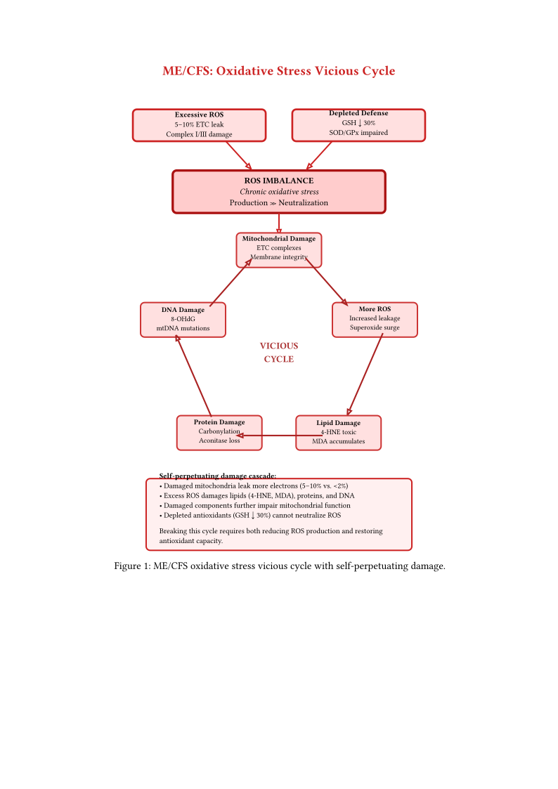
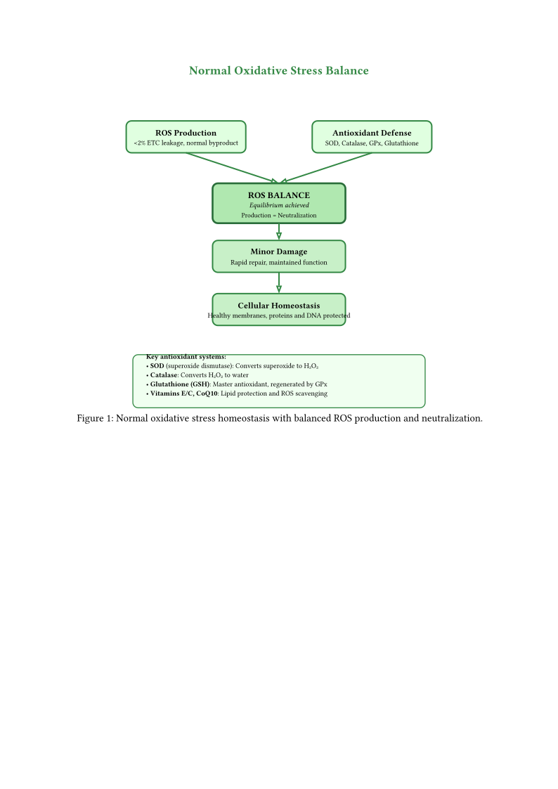
Figures oxidative stress normal and oxidative stress mecfs illustrate how oxidative stress creates a self-perpetuating cycle in ME/CFS, where excessive ROS production (5–10% electron leakage vs. normal 2%) combined with depleted antioxidants leads to progressive damage.
3 Oxidative and Nitrosative Stress in ME/CFS
Oxidative and nitrosative stress are consistently documented in ME/CFS and likely contribute to both mitochondrial dysfunction and symptom generation.
4 Reactive Oxygen Species (ROS)
4.1 Sources of ROS in ME/CFS
Multiple sources generate excess ROS:
- Mitochondrial electron leakage: Primary source during normal metabolism
- NADPH oxidase: Activated by immune stimulation
- Xanthine oxidase: Generates superoxide during purine metabolism
- Uncoupled eNOS: Produces superoxide instead of NO
- Inflammatory cells: Respiratory burst during immune activation
4.2 Damage to Cellular Components
ROS damage multiple targets:
- DNA: Base modifications, strand breaks, mutations
- Proteins: Carbonylation, cross-linking, loss of function
- Lipids: Peroxidation of membrane phospholipids
- Carbohydrates: Glycation reactions
4.3 Antioxidant System Dysfunction
The antioxidant defense system may be compromised:
- Glutathione: Often reduced in ME/CFS; critical for detoxification
- Superoxide dismutase (SOD): Variable findings
- Catalase: May be reduced
- Vitamins C and E: Nutritional antioxidants may be depleted
- Thioredoxin system: Important for protein redox balance
5 Reactive Nitrogen Species
5.1 Nitric Oxide Metabolism
Nitric oxide (NO) has complex roles in ME/CFS:
- Normal functions: Vasodilation, neurotransmission, immune defense
- iNOS induction: Inflammatory cytokines induce high NO production
- NO excess: Can inhibit mitochondrial respiration
- eNOS uncoupling: Produces superoxide instead of NO
5.2 Peroxynitrite Formation
When superoxide and NO react, they form peroxynitrite (ONOO-):
- Highly reactive: More damaging than either parent molecule
- Protein nitration: 3-nitrotyrosine formation (documented in ME/CFS)
- Lipid oxidation: Damages membrane integrity
- Mitochondrial inhibition: Irreversibly damages ETC complexes
5.3 Effects on Energy Metabolism
Nitrosative stress specifically impairs energy production:
- Complex I inhibition: NO reversibly inhibits electron transfer
- Complex IV inhibition: NO competitively inhibits oxygen binding at cytochrome c oxidase. Gut-derived hydrogen sulfide (H2S) inhibits the same CuB/hemea3~ site via a distinct mechanism (see h2s mitochondrial toxin) (Nicholls et al. 2013)
- Aconitase inactivation: Impairs Krebs cycle
- Glyceraldehyde-3-phosphate dehydrogenase: Inhibited by peroxynitrite
6 Lipid Peroxidation
6.1 Membrane Damage
Lipid peroxidation disrupts cellular membranes:
- Polyunsaturated fatty acids: Primary targets of peroxidation
- Chain reactions: One initiation event triggers multiple peroxidations
- Membrane fluidity: Peroxidation rigidifies membranes
- Permeability changes: Membranes become leaky
6.2 Isoprostanes and Other Markers
Lipid peroxidation products serve as biomarkers:
- F2-isoprostanes: Prostaglandin-like compounds from arachidonic acid peroxidation
- Malondialdehyde (MDA): End product of peroxidation
- 4-hydroxynonenal (4-HNE): Reactive aldehyde that modifies proteins
- Oxidized LDL: Marker of lipoprotein oxidation
Plasma F2-isoprostanes are elevated in ME/CFS across two independent studies. Kennedy et al. (\(n = 47\) CFS vs 34 controls) found elevated 8-iso-PGF2α; in a low-cardiovascular-risk subgroup, isoprostane levels correlated with PEM severity in a cross-sectional analysis (\(p = 0.027\), uncorrected, no effect size reported) (Kennedy et al. 2005). Robinson et al. (\(n = 33\) vs 33) independently confirmed elevation at rest, persisting 24 hours post-exercise (Robinson et al. 2010).
7 Research Gaps at Step 9
Six gaps at Step 9, all concerning the mitochondrial antioxidant machinery: (G33) PRDX3 expression in ME/CFS patient tissue (PRDX3-KO mice phenotypically resemble ME/CFS); (G34) NRF2 pathway activation status; (G35) thioredoxin / thioredoxin reductase (TRX/TXNRD) system; (G36) GPX4 lipid-peroxidation surveillance; (G37) direct muscle F2-isoprostanes (the gold-standard lipid peroxidation biomarker); (G38) mitochondrial glutathione (mGSH) pool.
| Gap | Essentiality | Worst-case impact | Tractability | Therapeutic leverage | Measurement | Priority |
|---|---|---|---|---|---|---|
| G33: PRDX3 expression | High — secondary H2O2 scavenger downstream of MnSOD; Prdx3-KO mice show reduced mtDNA content, reduced ATP, impaired mitochondrial fusion, and diminished exercise endurance (Lee 2020) (phenotype shared with many mitochondrial conditions, not ME/CFS-specific); Sonn et al. showed in cardiac tissue that PRDX3 protects PINK1 from OMA1-mediated degradation — PRDX3 loss impairs mitophagy (Sonn et al. 2022) (tissue context may differ in skeletal muscle) | Indirect: amplifies aconitase, Complex I, cardiolipin damage; additionally impairs mitophagy via PINK1 destabilization | Hard — muscle biopsy Western or IHC | Long-term | Muscle biopsy (Western or IHC) | 2 |
| G34: NRF2 pathway (master antioxidant regulator) | High — upregulates MnSOD, GPX1, catalase, thioredoxin, HO-1 simultaneously; but each target also has NRF2-independent regulation | Indirect but widespread; directionality unknown (suppressed vs compensatorily activated) | Moderate — NRF2 target mRNA panel in PBMCs (NQO1, HO-1, GCLC); not available through standard clinical labs | Research-stage — sulforaphane, dimethyl fumarate (requires lymphocyte monitoring), omaveloxolone (hepatic monitoring); premature pending NRF2 measurement | Blood draw (PBMC qPCR/RNA-seq; research lab, not standard clinical) | 1 |
| G35: Thioredoxin / TXNRD system | High — parallel to GSH as a major redox buffer; TXN transcript elevated in male Long COVID patients (Shankar 2025 ) but TXNRD1/2 protein and activity unmeasured | Indirect; could mask or worsen G33 | Moderate — erythrocyte TXNRD activity is available | Long-term | Blood draw (erythrocyte TXNRD activity) | 2 |
| G36: GPX4 lipid peroxidation surveillance | High — sole enzyme preventing ferroptotic membrane damage; GPX4 protein elevated 1.9× in ME/CFS lymphocytes (Shankar 2025, compensatory response) — but enzymatic activity, tissue-specific data (muscle/brain), and ferroptosis markers (ACSL4, 4-HNE) remain unmeasured | Indirect; links to cardiolipin peroxidation (Cardiolipin Peroxidation as the Convergence Point of ROS Damage) | Moderate — lymphocyte data exist; remaining work is activity assay and tissue-specific replication | Near-term — selenium, vitamin E, CoQ10 | Blood draw (PBMC data exist); muscle biopsy for tissue-specific | 1 |
| G37: Muscle F2-isoprostanes | Plasma F2-isoprostanes established as elevated in CFS: Kennedy 2005 (\(n = 47\) vs 34, correlated with PEM in low-cardiovascular-risk subgroup, \(p = 0.027\), cross-sectional) (Kennedy et al. 2005) and Robinson 2010 (\(n = 33\) vs 33, elevated at rest and persisting 24h post-exercise) (Robinson et al. 2010) — muscle-specific measurement remains genuinely open | Objective quantification of tissue-level ROS-damage state | Moderate — LC-MS assay available; muscle biopsy required for tissue-specific data | Immediate — baseline and response-to-intervention marker | Blood draw (plasma LC-MS); muscle biopsy for tissue-specific | 1 |
| G38: Mitochondrial glutathione pool (mGSH) | High — the cytosolic GSH compartment is not equivalent; mGSH is separately regulated | Direct on ROS scavenging capacity | Hard — requires subcellular fractionation from biopsy | Immediate — NAC, GSH precursors already in use | Muscle biopsy (subcellular fractionation) | 2 |
G34 (NRF2 pathway) and G37 (F2-isoprostanes) are priority 1 because they are tractable PBMC/plasma assays with immediately actionable interventions (sulforaphane, dimethyl fumarate; antioxidant dose-titration). G34 is notable because downstream NRF2 targets are documented as abnormal (Shankar 2025: reduced MnSOD protein, GPX4-mediated lipid peroxidation ), a review has identified NRF2 inhibition as a candidate contributor (Davis et al. 2025), yet the regulator itself has not been measured. The NRF2 target mRNA panel (NQO1, HO-1, GCLC) in PBMCs is a well-established assay requiring only a blood draw. The Long COVID bridge adds plausibility: Morris et al. showed that SARS-CoV-2 directly suppresses NRF2 protein and NRF2-dependent gene expression (Morris et al. 2023), raising the possibility that post-viral NRF2 suppression persists into ME/CFS. However, reduced downstream targets are also consistent with compensatory NRF2 upregulation overwhelmed by oxidative demand (see Step 9 discussion above); the measurement would resolve directionality. G36 (GPX4) is priority 1 because it connects directly to cardiolipin peroxidation (Cardiolipin Peroxidation as the Convergence Point of ROS Damage) and is already partially characterized by Shankar 2025 — the remaining work is replication. Step 9 as a whole is amplificatory rather than stoichiometric: its failure does not reduce per-cycle ATP output but accelerates damage to every other step, which is why antioxidant-targeted interventions have the potential for non-linear benefit once the primary ROS source is identified.
Certainty: 0.30 (conditional on itaconate shunt being active in ME/CFS, which itself remains unconfirmed; unconditional certainty is lower). Itaconate is a direct, potent NRF2 activator: 4-octyl-itaconate alkylates KEAP1 at Cys151, releasing NRF2 for nuclear translocation. If the metabolic safe mode model is correct and itaconate shunt activation is present in ME/CFS, then NRF2 should already be activated by itaconate-mediated KEAP1 modification. Yet downstream NRF2 targets are depleted (Shankar 2025: reduced MnSOD ). This creates a paradox: the problem may not be NRF2 suppression but rather NRF2 activation that is functionally overwhelmed by oxidative demand. Three sub-hypotheses: (a) NRF2 is nuclear and transcriptionally active, but antioxidant enzyme proteins are consumed faster than they can be synthesized; (b) NRF2 target gene promoters (NQO1, GCLC, HMOX1) are epigenetically silenced, preventing transcription despite nuclear NRF2; (c) chronic stress selects for NRF2 splice variants (e.g., NRF2-\(\Delta\)Neh2) with reduced transactivation capacity. These three interpretations have opposite therapeutic implications: if (a), adding NRF2 activators (sulforaphane) would be futile and the correct intervention is reducing oxidative demand (MitoQ, elamipretide); if (b), epigenetic modifiers would be needed; if (c), the NRF2 pathway itself requires isoform-specific targeting. The G34 measurement protocol should therefore include not just NRF2 protein and nuclear localization but also NRF2 target mRNA levels, promoter methylation status, and NRF2 isoform profiling.
This paradox connects to G39 (PGC-1\(\alpha\)) via the NRF2–PGC-1\(\alpha\) bidirectional circuit: PGC-1\(\alpha\) co-activates NRF2 transcription, and NRF2 induces PGC-1\(\alpha\) expression. In ME/CFS, both arms may be simultaneously impaired — PGC-1\(\alpha\) hyperacetylated (G39) and NRF2 overwhelmed (G34) — creating a broken hormetic loop. In healthy individuals, exercise generates a brief ROS pulse that activates NRF2, which cooperates with PGC-1\(\alpha\) to drive both antioxidant defense and mitochondrial biogenesis (mitohormesis). In ME/CFS, this loop is broken at every node: exercise is contraindicated (PEM), AMPK is impaired (Brown et al. 2018), PGC-1\(\alpha\) cannot be deacetylated (impaired SIRT1), and even if NRF2 is activated, the transcriptional programme is overwhelmed. The patient cannot exercise to activate hormesis, cannot activate PGC-1\(\alpha\) pharmacologically without AMPK, and the resulting antioxidant deficit makes any subsequent exercise more damaging — a self-reinforcing trap.
Testable prediction: NRF2 nuclear protein in ME/CFS PBMCs will be normal or elevated (not reduced); NQO1/HO-1 mRNA may be elevated; but the corresponding protein levels will be disproportionately low relative to mRNA (indicating post-transcriptional failure or excessive protein turnover). Falsified if NRF2 nuclear protein is genuinely reduced, consistent with simple suppression.