Cerebral Blood Flow Abnormalities
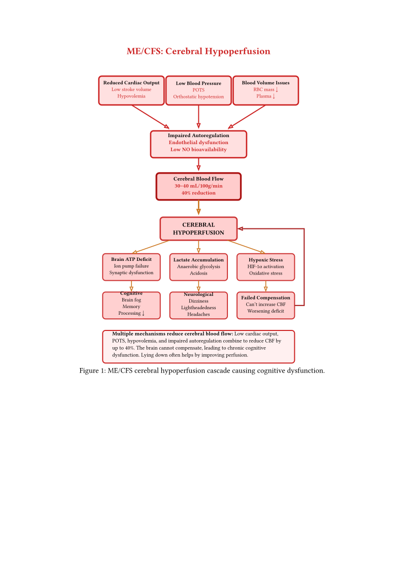
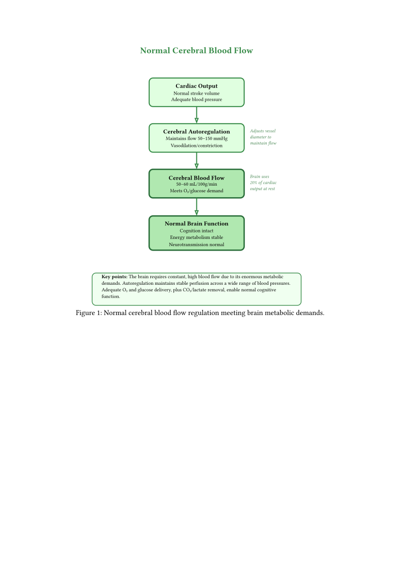
Cerebral blood flow (CBF) abnormalities are among the most consistently documented findings in ME/CFS and likely contribute substantially to cognitive symptoms.
Figures cerebral hypoperfusion normal and cerebral hypoperfusion mecfs illustrate how multiple mechanisms reduce cerebral blood flow in ME/CFS (30–40 mL/100g/min vs. normal 50–60 mL/100g/min, a 40% reduction).
1 Reduced Regional Blood Flow
Multiple neuroimaging modalities have demonstrated CBF reductions Campen, Rowe, and Visser (2020):
- Global hypoperfusion: 10–20% reduction in total cerebral blood flow (measured by SPECT and Doppler ultrasound)
- Regional deficits: Particularly in frontal, temporal, and parietal regions
- Brainstem hypoperfusion: Potentially explaining autonomic dysfunction (Barnden et al. 2011)
- Subcortical abnormalities: Basal ganglia and thalamic hypoperfusion
Van Campen et al. Campen, Rowe, and Visser (2020) documented that 90% of ME/CFS patients (n=429) showed abnormal CBF reduction (>13%) during head-up tilt testing, with end-tilt CBF reduction of 26% in ME/CFS patients versus only 7% in controls (n=44). Importantly, this occurred even in the absence of hypotension or tachycardia, indicating intrinsic cerebrovascular dysfunction rather than solely cardiovascular causes.
2 Correlation with Cognitive Symptoms
CBF reductions correlate with specific cognitive deficits:
- Frontal hypoperfusion → executive dysfunction, working memory impairment
- Temporal hypoperfusion → verbal memory deficits, language processing difficulties
- Parietal hypoperfusion → attention deficits, spatial processing impairment
- Global hypoperfusion → processing speed reduction, mental fatigue
3 Mechanisms of Cerebral Hypoperfusion
The cerebral hypoperfusion documented above likely results from multiple converging mechanisms:
- Reduced cardiac output: Secondary to autonomic dysfunction (Section Autonomic Nervous System Dysfunction) and blood volume deficits (Streeten and Bell 1998)
- Impaired cerebral autoregulation: Inability to maintain CBF across blood pressure changes (Barnden et al. 2011)
- Endothelial dysfunction: Reduced nitric oxide-mediated vasodilation
- Increased cerebrovascular resistance: Vasoconstriction or structural changes
- Neurovascular uncoupling: Failure of blood flow to match metabolic demand
The integration of autonomic dysfunction, reduced blood volume, and direct cerebrovascular pathology creates a multifactorial reduction in brain perfusion that correlates with cognitive symptom severity. This multifactorial integration is characteristic of ME/CFS pathophysiology and is discussed in the context of multi-system interactions in Chapter Integrative Models and Multi-System Pathophysiology, Section Multi-System Integration and Synthesis.
4 Exacerbation with Exertion
Importantly, cerebral perfusion abnormalities worsen following physical or cognitive exertion:
- Further CBF reductions post-exercise
- Prolonged recovery of normal perfusion
- Correlation with post-exertional malaise severity
- Potential contribution to cognitive “crashes” following activity
5 Cerebrovascular Reactivity to a Controlled Hypoxic Stressor
A proof-of-concept multimodal MRI study probed cerebrovascular reactivity by exposing ME/CFS patients (n=26) and controls (n=27) to normoxia plus two controlled hypoxic challenges (SpO2 ~87%), measuring whole-brain cerebral blood flow (CBF) with arterial spin labelling (Bader et al. 2026).
Certainty: 0.35. (Raw certainty 0.40 — Vienna preprint, not yet peer-reviewed, n=26/27; ME/CFS population weight 1.0 → discounted 0.40, then held at 0.35 because the variability finding is exploratory and unproven as a biomarker.) Whole-brain CBF rose under hypoxia in both groups (controls +4.8±13.0%, patients +3.7±11.7%), with no significant group-mean difference — gross cerebrovascular reactivity to a controlled hypoxic stressor was preserved on average (Bader et al. 2026) (Biswal, Kunwar, and Natelson 2011). However, at the first hypoxic challenge (H1), patient-to-control variance ratios were elevated across regions of interest (range 2.27–6.94), significant in three of four regions after false-discovery-rate correction — indicating greater inter-individual variability in the initial CBF response among patients (Bader et al. 2026) (He et al. 2013). These variance differences were absent at the second challenge (H2), suggesting either habituation or a transient dysregulation of the initial cerebrovascular adaptation rather than a fixed failure of reactivity.
Replication status: Not yet replicated. The variability finding is exploratory and derives from a single preprint.
Severity applicability: Unknown — the study cohort was not stratified by severity; whether this pattern holds across mild, moderate, severe, or very severe ME/CFS is not established.
Limitations: Proof-of-concept, modest sample, cross-sectional; the group-mean reactivity was preserved, so the claim rests on a variance-based, exploratory analysis; the hypoxic challenge is not physical exertion and cannot be linked to post-exertional malaise; whether variability in the initial response is a reproducible, severity-linked biomarker is unproven.
Falsifiable prediction: If the variability is a meaningful biomarker of dysregulated cerebrovascular adaptation, a second independent cohort should reproduce an elevated patient-to-control CBF-response variance ratio at an initial (unhabituated) challenge, and the degree of variability should correlate with symptom severity or orthostatic/cognitive load. Falsified if the variance difference does not reproduce, or if it reflects only methodological noise.
Consequence: This is an early, unproven clue that the difficulty may not be an average inability to change brain blood flow, but an unpredictable, person-to-person and moment-to-moment instability in that response — worth confirming, but not yet usable as a test or treatment target.
6 The Brainstem Autonomic Loop Hypothesis
The convergence of brainstem neuroinflammation (Yu et al. 2026 NII imaging (Yu2026diffusion?)-neuroinflammation), brainstem functional connectivity changes (Faro et al. 2024 (Faro et al. 2024)), cerebral hypoperfusion, and autonomic dysfunction suggests a three-node vicious cycle that may maintain ME/CFS pathology.
Certainty: 0.20. Downgraded from initial 0.40. Based on convergent neuroimaging (NII, fMRI), autonomic testing, and CSF proteomics evidence, but assembled from different studies in non-overlapping cohorts. Vicious cycle hypotheses are generically unfalsifiable (any intervention failure can be attributed to “targeting the wrong node”); the simpler alternative—all three nodes as parallel downstream consequences of a shared upstream event (e.g., post-viral endothelial damage, CNS energy deficit)—requires zero additional causal arrows. The vagal anti-inflammatory pathway, while established in animal models, is controversial in humans. (Yu2026diffusion?)-neuroinflammation (Faro et al. 2024) Bragée et al. (2026)
The brainstem (medulla, pons) contains autonomic centers—nucleus tractus solitarius, dorsal motor nucleus of vagus, rostral ventrolateral medulla—that regulate heart rate, blood pressure, and cerebral blood flow. Neuroinflammation in the brainstem could impair autonomic output, reducing vagal tone and the cholinergic anti-inflammatory pathway. Simultaneously, reduced autonomic output could produce hypovolemia and hypoperfusion, causing cerebral ischemia and metabolic stress that further drives neuroinflammation. However, the NII study shows widespread white matter changes, not brainstem-localized neuroinflammation; the Faro connectivity changes could reflect compensatory activation, disinhibition, or analysis artifact rather than autonomic failure; and the Bragee CSF evidence has no control group. Each of these three evidence pillars is weak individually, and treating them as measurements of three nodes in the same cycle in the same patients is an ecological fallacy.
Testable predictions: + NII metrics in brainstem white matter tracts will correlate more strongly with autonomic function tests (HRV, tilt table response) than with cognitive tests + POTS+ patients will show worse brainstem NII metrics than non-POTS patients + A shared upstream cause model predicts that all three nodes will correlate with a single third variable (e.g., overall disease severity); the cycle model predicts that each node predicts the others after controlling for severity