Hypothalamic-Pituitary-Adrenal (HPA) Axis
The hypothalamic-pituitary-adrenal axis represents one of the most extensively studied endocrine systems in ME/CFS, yet paradoxically remains among the most controversial. Through glucocorticoid signaling, the HPA axis coordinates the body’s stress response and regulates immune function while maintaining glucose homeostasis and energy metabolism. It further modulates circadian rhythms, sleep-wake cycles, cognitive function, and mood regulation. Given these critical roles, HPA dysfunction provides a plausible mechanism linking the diverse symptoms of ME/CFS.
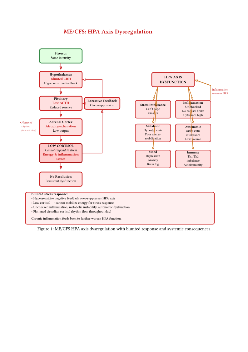
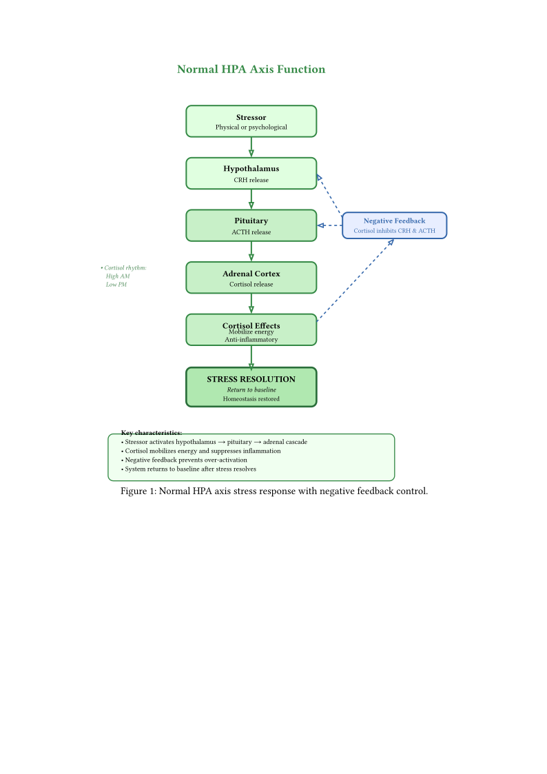
Figures hpa axis normal and hpa axis mecfs illustrate the characteristic pattern of HPA axis dysfunction observed in ME/CFS. Unlike the robust circadian cortisol rhythm and responsive feedback regulation seen in healthy individuals, ME/CFS patients demonstrate a distinct pattern of dysregulation. This involves blunted corticotropin-releasing hormone (CRH) secretion from the hypothalamus, reduced adrenocorticotropic hormone (ACTH) response from the pituitary, flattened diurnal cortisol rhythm with loss of the normal morning peak, and paradoxically enhanced negative feedback sensitivity. This constellation of abnormalities distinguishes ME/CFS from both healthy states and primary adrenal insufficiency (Addison’s disease), suggesting a unique form of central HPA axis hypofunction (Tomas et al. 2017) (Papadopoulos and Cleare 2012).
1 HPA Axis Abnormalities
1.1 Cortisol Dysregulation Patterns
Multiple studies document a consistent pattern of cortisol abnormalities in ME/CFS patients that differs qualitatively from both healthy individuals and patients with primary adrenal disorders. Baseline cortisol levels tend to be lower, though typically remaining within the broad “normal” laboratory reference range. The diurnal cortisol rhythm shows flattening, with reduced amplitude between morning and evening values. The cortisol awakening response (CAR)—the normal sharp rise in cortisol during the first 30–60 minutes after waking—appears attenuated. Additionally, cortisol responses to physiological and psychological stressors are blunted, despite appropriate ACTH response to exogenous CRH stimulation in some studies (Pipper et al. 2024). The NIH deep phenotyping study by Walitt et al. identified neuroendocrine abnormalities consistent with HPA axis dysfunction, including altered catecholamine metabolism that affects upstream regulation of the HPA axis (Walitt et al. 2024). The reduced central catecholamines documented in cerebrospinal fluid may contribute to impaired hypothalamic CRH release, providing a mechanistic link between neurological and endocrine dysfunction.
Certainty: 0.30. A single unpublished conference presentation (not yet peer-reviewed) from the University of Amsterdam (Netherlands Brain Bank ME/CFS programme), presented at the 2025 IACFS/ME conference, reports a pronounced reduction in CRH-immunoreactive neurons in the hypothalamic paraventricular nucleus of severe ME/CFS patients (\(n = 7\)) (Da Silva 2025). If confirmed, this would provide an anatomical substrate for the blunted central CRH drive and HPA hypoactivation documented in ME/CFS, relocating the origin of hypocortisolism from the adrenal gland to the hypothalamus. This finding, its cell-type specificity, its candidate drivers, and the substantial caveats attached to a single unpublished conference source (including whether it reflects neuron loss or peptide depletion) are developed in detail under the mechanisms of HPA dysfunction (Selective Depletion of Hypothalamic CRH Neurons in Severe ME/CFS).
Recent sex-stratified analysis by Pipper et al. (2024) revealed that cortisol dysregulation patterns differ significantly between male and female ME/CFS patients and vary by disease severity (Pipper et al. 2024). Female patients with severe ME/CFS demonstrated elevated 11-deoxycortisol (a cortisol precursor) and 17\(\alpha\)-hydroxyprogesterone, suggesting impaired final enzymatic steps in cortisol synthesis. Male patients with mild to moderate disease showed frankly reduced cortisol and corticosterone levels but paradoxically elevated progesterone. These findings indicate that HPA dysfunction may involve enzyme deficiencies in steroidogenesis rather than simple hypothalamic-pituitary signaling deficits.
1.2 ACTH and CRH Abnormalities
The central components of the HPA axis—CRH from the hypothalamus and ACTH from the pituitary—show complex abnormalities that do not fit simple models of endocrine failure. Studies employing CRH stimulation tests have yielded inconsistent results. Some report normal ACTH and cortisol responses to exogenous CRH administration, while others document blunted ACTH responses despite adequate CRH stimulation. Still others find normal ACTH responses but reduced cortisol output, suggesting adrenal hyposensitivity. These inconsistencies likely reflect the heterogeneity of ME/CFS patient populations, differences in disease duration and severity, and the limitations of single-timepoint testing to capture dynamic regulatory dysfunction.
The most consistent finding across studies is evidence of enhanced negative feedback sensitivity (Papadopoulos and Cleare 2012) (Tomas et al. 2017). Dexamethasone suppression tests demonstrate that low doses of synthetic glucocorticoid produce greater and more prolonged suppression of cortisol secretion in ME/CFS patients compared to controls. This suggests that the hypothalamus and pituitary remain exquisitely sensitive to glucocorticoid feedback signals, inappropriately dampening HPA axis output even when cortisol levels are already low-normal. This pattern resembles the neuroendocrine adaptation seen in chronic stress conditions but persists inappropriately in ME/CFS despite the clinical need for robust stress responses.
1.3 Diurnal Rhythm Disruption
The diurnal cortisol rhythm represents one of the most robust and well-characterized circadian processes in human physiology, yet ME/CFS patients consistently demonstrate flattening of this rhythm. Healthy individuals show a sharp cortisol peak within 30–60 minutes of waking (cortisol awakening response), followed by progressive decline throughout the day, reaching a nadir around midnight, and beginning to rise again in the early morning hours (3–4 AM) in anticipation of waking. In contrast, ME/CFS patients show reduced morning cortisol peak (blunted CAR), less pronounced decline during the day (flatter slope), and reduced overall amplitude (difference between peak and nadir), resulting in a “flattened” 24-hour pattern (Cambras et al. 2018). The mechanistic basis for circadian rhythm disruption extends beyond the HPA axis itself to involve the central circadian clock in the suprachiasmatic nucleus (SCN) of the hypothalamus. The NIH study documented abnormalities in temporal-parietal junction function and altered brain metabolism that may affect SCN regulation (Walitt et al. 2024). Additionally, inflammatory cytokines known to be elevated in ME/CFS (discussed in Chapter Immune System Dysfunction) directly disrupt circadian clock gene expression, creating bidirectional interactions between immune activation and circadian dysregulation.
The clinical consequences of flattened cortisol rhythm are profound. The morning cortisol peak serves essential physiological functions: promoting waking and alertness, mobilizing glucose for energy availability, preparing the cardiovascular system for upright posture and activity, and modulating immune function to prevent excessive inflammation. Loss of this peak explains the characteristic morning symptom severity reported by many ME/CFS patients. These include difficulty waking, prolonged morning fatigue requiring hours to achieve minimal function, orthostatic intolerance upon standing (discussed in Chapter Cardiovascular Dysfunction), and cognitive dysfunction particularly severe in early morning hours.
2 Mechanisms of HPA Dysfunction
The dysregulation of the HPA axis in ME/CFS reflects multiple interconnected mechanisms operating at different levels of the neuroendocrine cascade. Understanding these mechanisms is essential for developing targeted therapeutic interventions and explaining why simple hormone replacement strategies have shown limited efficacy.
2.1 Central Glucocorticoid Receptor Sensitivity
Falsifiability: weakly — Falsified if hypothalamic GR expression is normal or reduced in ME/CFS patients, or if elevated cortisol fails to suppress ACTH via enhanced feedback
The enhanced negative feedback sensitivity observed in ME/CFS may result from altered glucocorticoid receptor (GR) expression or function in hypothalamic and pituitary tissues. Several mechanisms could produce this effect. Upregulation of GR expression would increase sensitivity to existing cortisol levels. Altered GR isoform expression (GR\(\alpha\) vs. GR\(\beta\)) might shift the balance toward enhanced feedback. Reduced expression of 11\(\beta\)-hydroxysteroid dehydrogenase type 1 (11\(\beta\)-HSD1), the enzyme that locally amplifies cortisol action by converting inactive cortisone to active cortisol, could diminish local glucocorticoid signaling. Finally, epigenetic modifications of the GR gene might affect transcription and receptor function.
This enhanced feedback creates a self-reinforcing cycle. Slightly elevated cortisol (or even normal-low cortisol) triggers disproportionate suppression of CRH and ACTH secretion, further reducing cortisol output. Under normal circumstances, this would reduce feedback inhibition and restore output, but the hypersensitive feedback prevents this compensatory response, maintaining chronically low HPA axis activity. This mechanism explains why ME/CFS patients do not develop frank adrenal insufficiency (baseline cortisol remains detectable) yet fail to mount appropriate stress responses (blunted reactivity to challenges). This self-reinforcing HPA dysfunction represents one of several vicious cycles in ME/CFS pathophysiology, as discussed in Section Unifying Mechanisms Across Systems of Chapter Integrative Models and Multi-System Pathophysiology.
2.2 Inflammatory Cytokine Effects on HPA Axis
The bidirectional relationship between the immune system and the HPA axis represents a critical mechanism in ME/CFS pathophysiology. Under normal circumstances, immune activation from infection or tissue damage stimulates HPA axis activity. Pro-inflammatory cytokines (IL-1, IL-6, TNF-\(\alpha\)) signal the hypothalamus to increase CRH secretion, resulting in elevated cortisol that dampens the immune response. This creates negative feedback that prevents excessive inflammation. The acute response adaptively contains immune activation while preventing immunopathology.
Falsifiability: weakly — Falsified if ME/CFS immune cells show normal glucocorticoid receptor function and normal immune suppression at physiological cortisol concentrations
In ME/CFS, chronic low-grade inflammation (documented in Chapter Immune System Dysfunction) may induce glucocorticoid resistance at immune cells while simultaneously increasing central negative feedback sensitivity. This paradoxical pattern produces the worst of both scenarios: insufficient cortisol secretion to control peripheral inflammation due to enhanced central feedback, yet reduced cortisol effectiveness at immune cells due to receptor downregulation or dysfunction (Walitt et al. 2024) (Shahbaz et al. 2025). The result is persistent inflammation despite apparent “normal” cortisol levels that would typically suppress such immune activation.
Recent evidence from Shahbaz et al. (2025) documenting sex-specific immune dysregulation supports this model, showing that females with ME/CFS exhibit particularly pronounced pro-inflammatory profiles with elevated type 2 interferon signaling despite cortisol levels within the reference range (Shahbaz et al. 2025). This suggests functional glucocorticoid resistance at target tissues.
2.3 Steroidogenic Enzyme Dysfunction
The recent findings by Pipper et al. (2024) identifying elevated cortisol precursors (11-deoxycortisol, 17\(\alpha\)-hydroxyprogesterone) in severe ME/CFS patients suggest impaired function of steroidogenic enzymes, particularly 11\(\beta\)-hydroxylase (CYP11B1) which catalyzes the final step converting 11-deoxycortisol to cortisol (Pipper et al. 2024). This enzyme dysfunction could result from several factors. Mitochondrial impairment may play a role, as steroidogenesis occurs in mitochondria and requires adequate ATP supply (discussed in Chapter Energy Metabolism and Mitochondrial Function). Cytokine-mediated suppression of enzyme expression or activity represents another possibility. Micronutrient deficiencies affecting enzyme cofactors or oxidative stress damaging enzyme proteins may also contribute.
If confirmed, this mechanism suggests that the problem is not purely regulatory (hypothalamic-pituitary signaling) but also biosynthetic (adrenal enzymatic capacity). This has important therapeutic implications, as interventions targeting upstream signaling may prove ineffective if the limiting step is enzymatic conversion within the adrenal gland.
2.5 Structural Adrenal Atrophy
The mechanisms described above—enhanced glucocorticoid feedback, cytokine-mediated suppression, steroidogenic enzyme dysfunction, and post-viral pituitary injury—are primarily functional or signalling-level dysregulations. A fifth mechanism operates at the structural level: physical shrinkage of the adrenal glands themselves.
Scott and Dinan (1999) performed CT imaging of adrenal glands in 8 ME/CFS patients and healthy controls. All 8 patients had adrenal glands approximately half the volume of controls (Scott and Dinan 1999). All patients also showed lower 24-hour urinary free cortisol and blunted cortisol response to ACTH stimulation testing, consistent with reduced adrenal cortical mass limiting both hormone storage and synthesis capacity.
Study: (cross-sectional CT imaging, \(n = 8\) ME/CFS vs. controls, Psychoneuroendocrinology 1999; certainty: 0.45—striking finding but very small sample, not yet replicated).
The proposed mechanism is trophic: ACTH acts as a growth stimulus for the adrenal cortex. Chronically low ACTH output (from any of the upstream HPA dysfunction mechanisms) removes this trophic signal, leading to gradual adrenocortical atrophy over months to years. This creates a self-reinforcing vicious cycle: HPA dysfunction → low ACTH → adrenal atrophy → reduced cortisol capacity → inadequate stress responses → further HPA suppression via negative feedback.
This structural finding provides an important differential against major depression, where the HPA axis is hyperactive and adrenal glands enlarge due to chronic ACTH overstimulation. The opposite structural pattern in ME/CFS (hypoactive HPA, shrunken adrenals) argues against ME/CFS being a depressive disorder and supports a distinct neuroendocrine pathology.
The Scott and Dinan finding has not been independently replicated in 25+ years. The sample size (\(n = 8\)) precludes confident generalisation. Modern imaging studies with larger cohorts and correlation to illness duration, severity, and hormonal profiles are needed before structural atrophy can be considered an established feature of ME/CFS.
2.6 Central CRH-Neuron Loss in the Hypothalamus
The five mechanisms above locate HPA dysfunction at progressively more peripheral levels—enhanced central feedback, cytokine signalling, adrenal enzyme function, pituitary injury, and adrenal atrophy. A sixth candidate mechanism operates at the most upstream point of the entire axis: loss of the hypothalamic neurons that manufacture corticotropin-releasing hormone (CRH) itself. If the paraventricular nucleus (PVN) can no longer produce adequate CRH, every downstream compartment—pituitary ACTH, adrenal cortisol—is deprived of its initiating signal, and no intervention acting below the hypothalamus can restore normal regulation.
A preliminary brain-autopsy series presented at the IACFS/ME 2025 conference (Da Silva and colleagues, University of Amsterdam, using tissue from the Netherlands Brain Bank ME/CFS donation programme) reported that the hypothalamus of seven severely affected, deceased ME/CFS patients contained dramatically reduced numbers of CRH-immunoreactive neurons in the PVN—in some cases almost none—compared with matched controls (Da Silva 2025). Because these counts reflect CRH-peptide immunostaining, they cannot by themselves distinguish loss of neurons from loss of detectable peptide in surviving cells (see limitation below). The change was reported as cell-type specific: neurons producing vasopressin (AVP) and oxytocin (OXT) were normal or relatively preserved, arguing against a global hypothalamic atrophy. Downstream, the pituitary reportedly showed downregulated receptors for regulatory hormones and reduced output of the ACTH precursor pro-opiomelanocortin (POMC). If borne out, this pattern would relocate the origin of the well-documented ME/CFS hypocortisolism (Hypothalamic-Pituitary-Adrenal (HPA) Axis; meta-analytic hypocortisolism (Tak et al. 2011)) from the adrenal gland to the hypothalamus, reframing low morning cortisol as the readout of a central manufacturing deficit rather than an adrenal or feedback problem.
The single most important caveat is evidential: this is an unpublished conference presentation (\(n = 7\), severe/very-severe cases only, single centre, no peer review or independent replication as of 2026). It is reported here at low certainty and must not be treated as an established feature of ME/CFS. The finding is nonetheless notable because direct human hypothalamic histology in ME/CFS is exceptionally rare—the prior peer-reviewed autopsy literature is limited to isolated reports of gliosis and white-matter change without cell-type-specific hypothalamic quantification (Ferrero et al. 2017).
Certainty: 0.30 — single non-peer-reviewed source; strong methodological lineage (the Swaab group’s PVN CRH-neuron quantification is well established (Bao and Swaab 2010) (Bao, Meynen, and Swaab 2008)) but no ME/CFS-specific replication.
Refuted if: independent, blinded stereological quantification of the PVN finds CRH-neuron numbers in severe ME/CFS comparable to matched controls, or the apparent deficit disappears after controlling for agonal state and medication.
Consequence: If replicated, this would move the presumed root of the stress-hormone failure in severe ME/CFS from the adrenal gland up to the brain itself, meaning that cortisol deficiency would be secondary to lost central drive rather than a primary adrenal or feedback problem—but the finding is far too preliminary to change any clinical decision today.
The reported direction of the CRH change is as informative as its magnitude. In major depression (Raadsheer et al. 1994) and in multiple sclerosis (Purba et al. 1995) (both studied by the same Netherlands Brain Bank methodology) PVN CRH-expressing neurons are increased, reflecting a hyperactive or up-driven stress axis. Da Silva’s ME/CFS finding is the opposite: CRH-immunoreactive neurons reduced. This reversal, if confirmed, would raise questions about whether ME/CFS hypocortisolism is the tail end of chronic stress-system overdrive (“burnout”)—which would predict CRH-neuron preservation or increase—and would instead be consistent with a distinct process that selectively silences or depletes peptide from CRH neurons while sparing neighbouring AVP and OXT populations. A cell-type-specific vulnerability of this kind would parallel the selective GnRH-neuron death documented in the hypothalamus of Long COVID patients (Sauve et al. 2023) (a cross-disease precedent, not yet demonstrated in ME/CFS), consistent with a shared post-infectious mechanism that targets particular hypothalamic neuronal subtypes rather than the region as a whole.
Certainty: 0.25 — rests on the same unpublished finding plus cross-condition histology; the ME/CFS-versus-depression contrast is indirect (different cohorts, not a head-to-head study).
Refuted if: a single-run blinded stereology study finds ME/CFS PVN CRH-neuron counts at or above control/depression levels, or finds AVP/OXT neurons also depleted (indicating global rather than cell-type-selective loss).
Consequence: If the pattern holds, it would give ME/CFS a biological signature that is the mirror image of depression at the level of brain tissue—further evidence that the two illnesses are mechanistically different, even where their surface symptoms overlap.
The mechanism that would produce selective CRH-neuron depletion is unresolved, and the candidate drivers make competing, testable predictions.
- Chronic neuroinflammation / excitotoxicity:: Years of low-grade inflammatory or excitatory overactivation of stress centres could progressively damage the metabolically demanding CRH neurons. This is consistent with immune-inflammatory models of central HPA hypofunction (Morris, Anderson, and Maes 2017). However, it is constrained by a null finding: TSPO-PET studies have not consistently detected neuroinflammation in ME/CFS, though TSPO imaging has limited sensitivity for chronic low-grade hypothalamic inflammation and attributes signal ambiguously across cell types.
- Autoimmune targeting:: Anti-pituitary and anti-hypothalamus autoantibodies have been detected in a subset of CFS patients (De Bellis et al. 2021), offering an alternative in which CRH-producing cells (or their pituitary targets) are damaged by autoimmunity rather than inflammation. Distinguishing this from the inflammatory model requires simultaneous autoantibody and histological data in the same brains—which do not yet exist.
- Post-infectious neuronal death:: The GnRH-neuron precedent (Sauve et al. 2023) raises the possibility of direct viral or para-infectious injury to specific hypothalamic subtypes.
These are not mutually exclusive, and the current evidence cannot adjudicate between them. The decisive experiment is multiplexed staining of the same CRH-depleted PVN sections for activated microglia, T-cells, and IgG deposition: microglial nodules concentrated in depleted zones would support the inflammatory model, IgG selectively on CRH (not AVP/OXT) neurons would support the autoimmune model, and the absence of both despite CRH loss would point to a metabolic/excitotoxic driver.
Consequence: Which driver is correct matters for treatment: an inflammatory cause would point toward anti-inflammatory or immune-calming therapies, an autoimmune cause toward immunomodulation, and neither would be helped by simply replacing the missing hormone—so resolving this question is a prerequisite for any rational therapy aimed at the cause.
If PVN CRH neurons are genuinely depleted, the loss should have consequences beyond low cortisol, because these neurons contribute to two circuits relevant to core ME/CFS symptoms. (Origin: brainstorm.)
- Loss of central stress-induced analgesia:: CRH and the related urocortins act as endogenous analgesics through CRH receptors in the periaqueductal grey, rostral ventromedial medulla, and spinal dorsal horn. Reduced output from the principal central CRH source would weaken this tonic inhibition of pain transmission, predicting reduced stress-induced analgesia and raised baseline pain sensitivity—a plausible contributor to the widespread pain and fibromyalgia overlap seen in ME/CFS, framed as an abnormal stress–pain interaction rather than simply “more pain.”
- Loss of exertion-evoked sympathetic reserve:: Parvocellular PVN CRH neurons project to brainstem presympathetic centres and shape the acute sympathetic response to stressors. Selective loss (with AVP/OXT and baseline autonomic tone spared) predicts a reserve-dependent deficit: normal resting heart rate and blood pressure but impaired orthostatic and thermoregulatory compensation specifically when demand rises, consistent with the documented exertional and orthostatic intolerance of ME/CFS.
Both predictions are indirect—no ME/CFS study has measured CRH-neuron number against pain or autonomic reserve—and each rests on the unconfirmed depletion finding, assumes permanent circuit loss rather than recoverable peptide depletion, and assumes PVN CRH is the dominant functional pool (extrahypothalamic CRH sources such as the central amygdala and bed nucleus of the stria terminalis could partially compensate).
Certainty: 0.18 — physiologically grounded but doubly contingent (on the depletion finding and on untested ME/CFS-specific links).
Refuted if: ME/CFS patients show normal conditioned pain modulation and normal stress-evoked sympathetic responses (with intact orthostatic/thermoregulatory compensation on challenge), or these measures show no relationship to HPA-axis output.
Consequence: If correct, some of the pain and the activity-triggered crashes in ME/CFS would trace back to the same small population of lost brain cells—but this is a reasoned prediction awaiting the basic experiments to test it, not a current explanation.
Because the finding is preliminary, its value lies chiefly in the specific, feasible experiments it invites—most executable on existing Netherlands Brain Bank tissue. (Origin: brainstorm.)
- Resolve the driver:: Multiplexed staining of the same PVN sections for CRH neurons, activated microglia (IBA1/CD68), T-cells (CD3), and human IgG would show whether CRH-depleted zones colocalise with inflammatory infiltrate, autoantibody deposition, or neither—distinguishing the neuroinflammatory, autoimmune, and metabolic/excitotoxic models in one experiment.
- Test the depression contrast directly:: A single blinded stereology run comparing severe ME/CFS, severe major depression, and matched controls in one staining batch would establish whether the reported ME/CFS decrease versus the known depression increase (Raadsheer et al. 1994) (Purba et al. 1995) is a real biological reversal or a cross-study artefact.
- Distinguish cause from consequence:: Regressing CRH-neuron count on documented disease duration tests whether loss is progressive (ongoing, implying a treatment window) or a fixed early deficit.
- Look beyond CRH:: Single-nucleus RNA sequencing of hypothalamic micropunches would reveal whether other neuron subtypes are also affected and would carry a glial transcriptomic signature indicating the dominant pathological process.
Consequence: The finding’s main present value is that it points to a short list of concrete experiments, most executable on existing Netherlands Brain Bank tissue, that could within a small number of studies either establish a brain-tissue signature of ME/CFS or retire the claim.
Two further possibilities are recorded for completeness at very low certainty. (Origin: brainstorm.) First, by analogy to the selective GnRH-neuron death seen after SARS-CoV-2 (Sauve et al. 2023), CRH-neuron loss might be trigger-specific—present only in ME/CFS following particular neurotropic infections and absent in other onset types, which would make it a subtype marker rather than a universal feature. Second, the autoimmune and neurodegenerative accounts might be unified in a two-hit model in which anti-hypothalamus autoantibodies (De Bellis et al. 2021) supply targeting specificity (explaining why CRH but not AVP/OXT neurons are affected) while complement and microglia supply the effector killing. Both ideas are currently too weakly evidenced for independent development and are retained only as directions for future cycles should the core finding replicate.
Certainty: 0.10 — speculative extensions of an already-preliminary finding.
Refuted if: CRH-neuron loss proves trigger-independent across onset types, or PVN co-staining shows no IgG deposition on CRH neurons.
Consequence: These are placeholders for future investigation, not claims—if the basic finding fails to replicate, both fall away.
Several boundaries constrain any use of the CRH-depletion finding. It derives from a single unpublished conference presentation with no primary publication or independent replication as of 2026, reported only secondarily; “near-absent” may be a paraphrase rather than the investigators’ quantitative language. The quantification method is undisclosed—if it was qualitative or unblinded rather than the Swaab group’s unbiased stereology (Bao, Meynen, and Swaab 2008), observer bias aligned with the well-known expectation of HPA dysfunction in ME/CFS is a real risk. A specific technical confound also applies: CRH is a fast-turnover neuropeptide, so terminal HPA hyperactivation could deplete peptide stores below the immunostaining threshold in structurally intact neurons, making “near-absent CRH staining” potentially reflect peptide exhaustion rather than cell death (resolvable by CRH mRNA in-situ hybridisation). The sample (\(n = 7\)) comprised only severe and very-severe deceased patients; whether comparable CRH-neuron loss occurs in mild or moderate ME/CFS is entirely unknown, and it may represent an end-stage change—or a consequence of prolonged immobility, cachexia, polypharmacy, agonal state, or post-mortem interval—rather than an ME/CFS-specific feature, absent comparison to non-ME/CFS chronically-ill controls. As end-of-life tissue, it cannot distinguish whether CRH-neuron loss is a cause of ME/CFS, a consequence of severe illness, or an artefact. No CRH quantification in other brain regions was reported, so region-specificity is unconfirmed. Until a peer-reviewed publication with a larger, severity-stratified cohort and matched controls appears, this mechanism should be treated as a provocative hypothesis-generating observation, not an established mechanism.
Consequence: This finding is exciting but not yet trustworthy enough to act on—patients and clinicians should regard it as a promising lead to watch, not a reason to change diagnosis or treatment.
DHEA deficiency prevalence: Nathan (2013) (Nathan 2013), drawing on clinical experience with over 5,000 patients, reported that more than 90% of fibromyalgia and ME/CFS patients are DHEA-deficient, “often to a profound degree.” DHEA supplementation has shown promising results in ME/CFS (see Section on Testosterone and Androgens below for details). Interpretation of DHEA results requires age-appropriate reference ranges, as levels naturally decline from a peak in adolescence; a result appearing “normal” on wide laboratory ranges may represent functional deficiency for a younger patient.
3 Clinical Consequences
The HPA axis abnormalities documented in ME/CFS produce wide-ranging clinical effects that contribute directly to the cardinal symptoms of the disease. Understanding these consequences illuminates why seemingly minor hormonal changes cause profound functional impairment.
3.1 Stress Response Abnormalities and Post-Exertional Malaise
ME/CFS patients demonstrate inadequate cortisol responses to physiological stressors. These include exercise (both acute bouts and prolonged exertion), orthostatic challenge (standing, tilt-table testing), cognitive tasks requiring sustained mental effort, and psychological stressors. This blunted response means that stressors that healthy individuals accommodate with transient cortisol elevation produce inadequate counter-regulatory responses in ME/CFS patients, potentially explaining the delayed and prolonged symptom exacerbation characteristic of post-exertional malaise (PEM). The temporal pattern of PEM—symptom onset typically 12–48 hours after exertion rather than immediately—aligns with the kinetics of cortisol’s effects on immune function and cellular metabolism. During exertion, ME/CFS patients may rely on sympathetic nervous system activation (catecholamines) to maintain function despite inadequate cortisol support. Following exertion, the delayed cortisol response fails to adequately suppress the inflammatory cascade initiated by exertion-induced cellular stress and damage. This unchecked inflammation then drives the delayed symptom exacerbation of PEM.
3.2 Immune System Effects and Chronic Inflammation
Cortisol serves as the body’s primary endogenous anti-inflammatory hormone. It suppresses pro-inflammatory cytokine production (IL-1\(\beta\), IL-6, TNF-\(\alpha\)), inhibits T cell activation and proliferation, promotes a shift from Th1 (cellular immunity) to Th2 (humoral immunity) responses, and prevents autoimmune reactions by maintaining immune tolerance. The blunted cortisol output and flattened diurnal rhythm in ME/CFS remove this tonic immunosuppressive influence, permitting chronic low-grade inflammation to persist.
Falsifiability: weakly — Falsified if restoring the morning cortisol peak through timed hydrocortisone replacement fails to reduce inflammatory markers across the circadian cycle
The flattened cortisol rhythm may be particularly consequential for immune regulation. Immune cells express glucocorticoid receptors and show circadian variation in their responsiveness to cortisol. The normal morning cortisol peak serves to “reset” immune function daily, preventing inflammatory pathways from remaining chronically activated. Loss of this peak in ME/CFS may allow inflammatory signaling to persist across day-night cycles without the normal circadian suppression (Cambras et al. 2018).
This mechanism connects to the findings in Chapter Immune System Dysfunction documenting altered cytokine profiles, NK cell dysfunction, and B cell abnormalities in ME/CFS. The sex-specific immune profiles identified by Shahbaz et al. (2025) showing more pronounced inflammatory signatures in females align with the sex-specific steroid hormone abnormalities documented by Pipper et al. (2024), suggesting coordinated sex-dependent endocrine-immune interactions (Shahbaz et al. 2025) (Pipper et al. 2024).
3.3 Energy Metabolism and Glucose Homeostasis
Cortisol plays essential roles in energy metabolism. It stimulates hepatic gluconeogenesis to maintain blood glucose availability, promotes lipolysis (fat breakdown) to provide alternative fuel sources, modulates insulin sensitivity to optimize glucose utilization, and supports mitochondrial function and cellular energy production. HPA axis dysfunction directly impairs these metabolic processes.
The cerebral glucose hypometabolism documented by PET imaging studies (Tirelli et al., 1998; Siessmeier et al., 2003) may partly reflect inadequate cortisol support for glucose uptake and utilization (Tirelli et al. 1998) (Siessmeier et al. 2003). Additionally, the blunted morning cortisol peak fails to provide the metabolic “boost” needed to transition from fasting metabolism to active daytime metabolism, contributing to severe morning fatigue and the prolonged time required to achieve minimal function after waking.
These metabolic effects connect directly to the mitochondrial dysfunction and energy metabolism deficits discussed in Chapter Energy Metabolism and Mitochondrial Function, suggesting that HPA axis abnormalities and cellular metabolic dysfunction represent interconnected rather than independent pathophysiological processes.
Gut Barrier Repair and Low Cortisol in Severe Patients.
Beyond the well-established role of cortisol in stress response and metabolism, emerging evidence suggests that HPA axis dysfunction in severe ME/CFS may impair intestinal barrier maintenance and repair capacity, potentially contributing to chronic gut permeability and systemic inflammation.
3.4 Low Morning Cortisol in Severely Ill Patients.
A comprehensive biomarker examination of severely ill (housebound/bedbound) ME/CFS patients (Komaroff and Lipkin 2021) documented significantly reduced morning salivary cortisol compared to age- and sex-matched healthy controls: median 0.20 mcg/dL in severe ME/CFS vs. 0.45 mcg/dL in controls (p = 0.002). To contextualize these values clinically: normal morning salivary cortisol ranges from approximately 0.10 to 0.90 mcg/dL, with 0.20 mcg/dL representing roughly the 15th percentile and 0.45 mcg/dL representing the 50th percentile of the healthy population distribution. Thus, severe ME/CFS patients exhibit cortisol levels that, while not meeting formal diagnostic criteria for adrenal insufficiency (\(<\) 0.10 mcg/dL), fall well below the physiological optimum for barrier maintenance and metabolic function. This 55% reduction in morning cortisol suggests substantial HPA axis dysregulation in the severe patient population. While the mechanisms remain debated, the functional consequence is reduced cortisol availability during periods when barrier repair processes are most active.
3.5 Cortisol’s Role in Intestinal Barrier Function.
Glucocorticoids, including cortisol, play multifaceted roles in maintaining epithelial barrier integrity: (1) upregulating tight junction proteins (claudin-1, occludin, ZO-1) through glucocorticoid receptor-mediated transcription; (2) suppressing barrier-disrupting cytokines (IL-1\(\beta\), IL-6, TNF-\(\alpha\)) via NF-\(\kappa\)B inhibition; (3) promoting enterocyte survival and differentiation; and (4) maintaining circadian tight junction protein expression with peak synthesis during the morning cortisol surge. The relationship is dose-dependent: physiological cortisol levels are barrier-protective, while low cortisol impairs repair capacity. ME/CFS patients appear to have insufficient cortisol for normal barrier maintenance.
3.6 Impaired Barrier Repair in Severe ME/CFS: Mechanistic Hypothesis.
Integrating low cortisol with evidence of baseline gut permeability (Martín-Núñez et al. 2023), severe patients likely exhibit: (1) daily micro-damage from minimal activities (cognitive work, postural changes, meals) triggering transient splanchnic hypoperfusion; (2) insufficient nocturnal cortisol for barrier repair; (3) accumulating baseline permeability; (4) the cytokine-barrier bidirectional cycle (cytokines → tight junction disruption → LPS → cytokine amplification); and (5) nutritional deficits (low albumin in severe patients) limiting epithelial regeneration substrate. This model suggests wheat elimination response may be slower in severe patients due to impaired barrier repair capacity, but nutritional support (protein, micronutrients) and optimization of morning cortisol timing may accelerate recovery.