Adaptive Immunity
The adaptive immune system provides specific, long-lasting responses through T and B lymphocytes. The NIH deep phenotyping study identified characteristic abnormalities in B cell populations that may represent a biomarker signature for ME/CFS (Walitt et al. 2024).
1 T Cell Abnormalities
T lymphocytes coordinate adaptive immune responses and directly eliminate infected cells.
1.1 T Cell Subset Distribution
CD4/CD8 Ratio Changes The ratio of helper (CD4+) to cytotoxic (CD8+) T cells is altered in some ME/CFS patients, though findings vary considerably across studies . Some report a decreased CD4/CD8 ratio while others find an increased ratio. This heterogeneity may reflect distinct patient subgroups within the ME/CFS population.
Helper T Cell Subsets CD4+ T cells differentiate into functional subsets with distinct roles: Th1 cells produce interferon-gamma and promote cell-mediated immunity; Th2 cells produce IL-4, IL-5, and IL-13 to promote antibody responses; Th17 cells produce IL-17 and are involved in autoimmunity and mucosal defense; and regulatory T cells (Tregs) suppress immune responses to maintain tolerance. ME/CFS findings include Th1/Th2 imbalance (though the direction varies across studies), elevated Th17 cells in some patients, and reduced Treg numbers or function (Ekua W. Brenu et al. 2014). Altered cytokine profiles reflect these subset imbalances.
1.2 T Cell Exhaustion Markers
Chronic antigen exposure can lead to T cell exhaustion, characterized by:
- Increased PD-1 expression: Programmed death-1, an inhibitory receptor (Derek S. Iu et al. 2024) (Walitt et al. 2024)
- Elevated Tim-3: T cell immunoglobulin and mucin domain-3 (Derek S. Iu et al. 2024)
- CTLA-4 upregulation: Cytotoxic T-lymphocyte-associated protein 4 (Derek S. Iu et al. 2024)
- Reduced proliferative capacity: Impaired response to stimulation (Derek S. Iu et al. 2024)
- Decreased cytokine production: Despite activation marker expression (Derek S. Iu et al. 2024)
These findings suggest chronic immune stimulation in ME/CFS, consistent with persistent infection or autoimmune processes (Derek S. Iu et al. 2024).
Comprehensive T Cell Exhaustion Evidence (Iu et al. 2024)
A 2024 study published in PNAS provided the most detailed characterization of T cell exhaustion in ME/CFS to date (Derek S. Iu et al. 2024). Using transcriptomic and epigenetic profiling, Iu et al. demonstrated that CD8+ T cells from ME/CFS patients undergo extensive reprogramming toward an exhausted phenotype.
1.3 Key Findings
- Elevated PD-1 expression: Confirmed at both protein and transcriptional levels
- Transcriptional reprogramming: Gene expression patterns characteristic of chronic antigenic stimulation
- Epigenetic modifications: Persistent chromatin changes indicating long-term immune activation rather than transient response
- Similarity to chronic infections: The exhaustion profile resembled that seen in chronic viral infections (HIV, hepatitis C) and cancer
1.4 Implications
The epigenetic nature of these changes suggests that T cell exhaustion in ME/CFS is not merely a snapshot of current immune activation but represents a durable reprogramming of immune cell function. This has several implications:
- Chronicity: The epigenetic changes may explain why immune dysfunction persists even if the initial trigger resolves
- Impaired viral control: Exhausted T cells cannot effectively clear viruses, potentially permitting herpesvirus reactivation
- Therapeutic targets: Immune checkpoint inhibitors (anti-PD-1, anti-CTLA-4) used in cancer might theoretically restore T cell function, though safety in ME/CFS is unknown
- Biomarker potential: T cell exhaustion markers could serve as diagnostic or prognostic indicators
1.5 Integration with NIH Deep Phenotyping Study
The Iu et al. findings complement the Walitt et al. NIH study (Walitt et al. 2024), which also documented elevated CD8+ T cell PD-1 expression. Together, these studies consistently document T cell exhaustion as a reproducible feature of ME/CFS immunopathology, supporting the model of chronic antigenic stimulation driving both B cell (naïve/memory imbalance) and T cell (exhaustion) abnormalities.
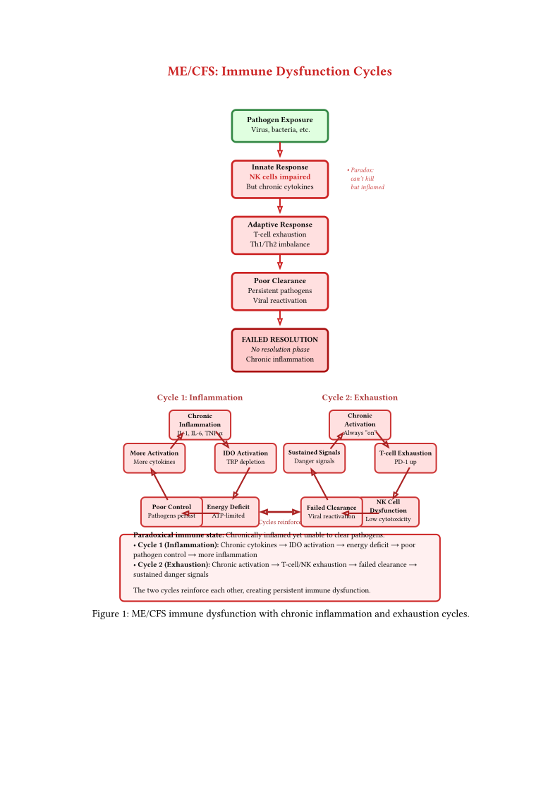
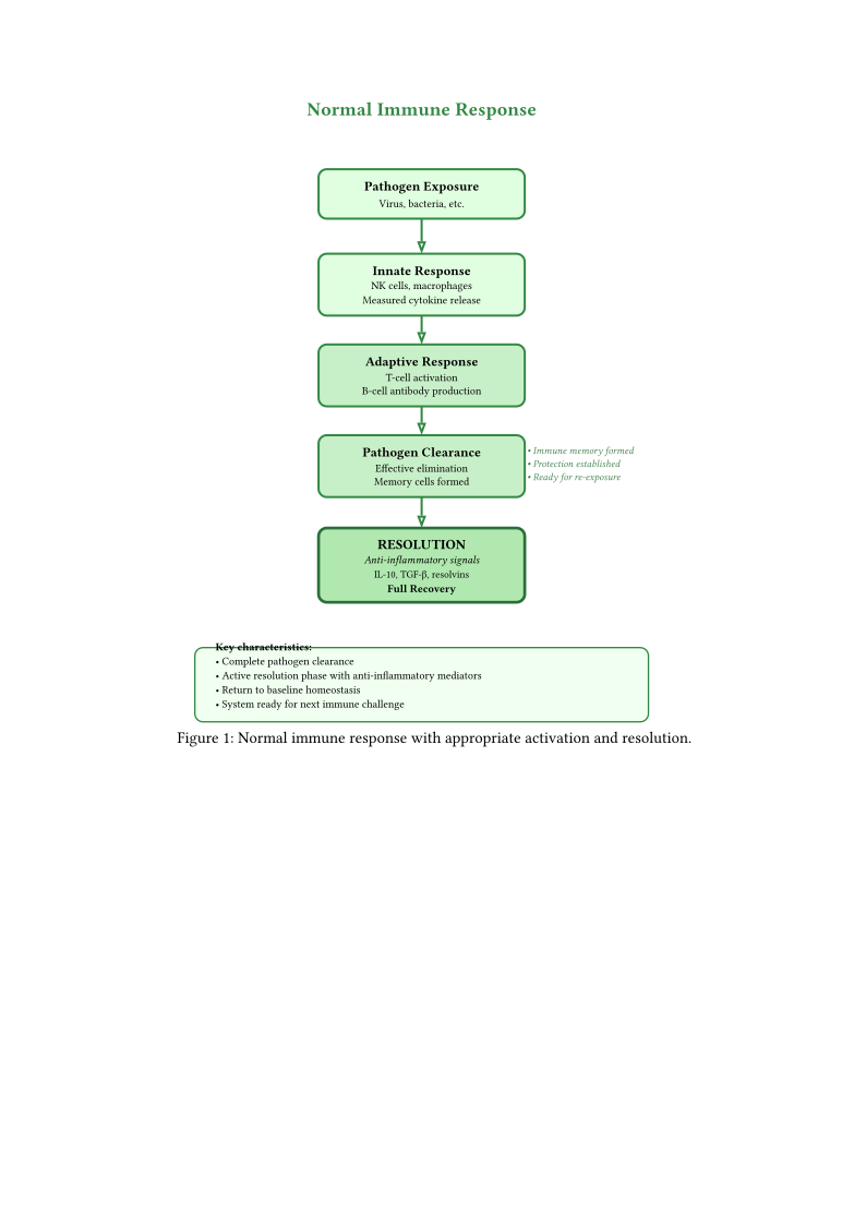
Figures immune normal and immune mecfs illustrate the paradoxical immune state in ME/CFS—simultaneously overactive and underactive. Two interconnected vicious cycles drive disease: chronic inflammation (IDO activation, energy deficit, poor pathogen control) and immune exhaustion (T-cell/NK dysfunction, failed clearance). These cycles reinforce each other. The integration of these immune-specific vicious cycles with metabolic and autonomic cycles is examined in Section Unifying Mechanisms Across Systems of Chapter Integrative Models and Multi-System Pathophysiology.
T cell exhaustion markers (PD-1, Tim-3, CTLA-4) in ME/CFS resemble those seen in chronic viral infections and cancer. However, the antigenic source driving this exhaustion has not been identified. The exhaustion profile is consistent with chronic antigenic stimulation but does not establish what the antigen is (persistent virus, autoantigen, or metabolic danger signal). Without identifying the driver, the therapeutic implication that checkpoint inhibitors might help remains speculative and carries substantial autoimmune risk.
(Certainty: 0.45 — T-cell exhaustion documented at epigenetic level in a single scRNA-seq study (n = 28/30; unreplicated); EV-cargo mechanism remains mechanistic inference from Ewing sarcoma.)
CD8+ T-cell exhaustion in ME/CFS has been documented in a single unreplicated scRNA-seq study. Iu et al. showed by scRNA-seq and ATAC-seq (n = 28 ME/CFS, 30 controls) that CD8+ effector memory T cells carry epigenetically locked exhaustion programs: TBX21/EOMES transcription factor upregulation, TCF7 chromatin closure (the hallmark of terminal exhaustion), and reduced cytotoxic gene expression. Critically, PD-L1/2 and CD86 were upregulated on ME/CFS monocytes — pointing to an immunosuppressive myeloid cell as the immediate suppressor, consistent with the MDSC hypothesis MDSC Expansion via Exosomal HSAT2 as the Upstream Cause of NK Cytotoxicity Loss. Eaton-Fitch et al. confirmed shared PD-1/CTLA-4 checkpoint gene dysregulation in both ME/CFS and Long COVID (n = 14/15/18), with ME/CFS showing the more suppressed and Long COVID the more activated exhaustion phenotype.
Standard accounts assume chronic antigenic stimulation as the upstream driver, but no consistent antigen has been identified in ME/CFS. An alternative mechanism emerges from Evdokimova et al. : exosomal uptake of HSAT2/HERV-K RNA by CD8+ T-cells directly induces the exhaustion phenotype (CD8+CD25+PD-1+) independently of antigen presentation. The PD-L1 upregulation on monocytes observed by Iu et al. is mechanistically consistent with an MDSC-like myeloid cell driving checkpoint-mediated T-cell suppression. This would reframe PD-1 upregulation and exhaustion in ME/CFS as a consequence of exosomal cargo programming rather than a marker of chronic antigen exposure.
This matters therapeutically: if exhaustion is antigen-driven, the antigen must be eliminated to reverse it. If exhaustion is EV-cargo-driven, removing or blocking the offending EVs could restore T-cell function without requiring pathogen identification. Checkpoint inhibitors (anti-PD-1) would provide temporary symptomatic relief at best, with exhaustion returning as long as the exosomal loop persists.
Falsifiable prediction: Plasma EV HSAT2 levels will correlate with CD8+ T-cell PD-1 expression longitudinally within individuals (within-subject r > 0.5 across ≥ 6 timepoints). EV depletion from patient plasma (size-exclusion filtration) before incubation with healthy CD8+ T-cells will abolish or reduce the PD-1 induction compared to unfiltered plasma. If PD-1 induction requires antigen-presenting cells rather than direct EV uptake, antigen-driven exhaustion remains more likely. The epigenetic TCF7 closure (Iu 2024) could be tested for reversibility by EV removal.
Limitations: The EV-cargo mechanism was demonstrated in vitro with high EV concentrations from Ewing sarcoma cells; whether chronic low-level ME/CFS EVs produce the same phenotype is unknown. The Iu 2024 PD-L1 finding is on monocytes, not specifically CD33+HLA-DR− MDSCs. The antigen-driven and EV-cargo-driven models are not mutually exclusive — both could contribute. Replication: exhaustion confirmed (Iu 2024, Eaton-Fitch 2024); EV mechanism not replicated in any chronic disease context.
(Certainty: 0.40 — phenotypic overlap is mechanistically plausible; direct MDSC gating of the Iu 2024 cohort not yet performed.)
Iu et al. identified PD-L1/2-upregulated monocytes as the proximate suppressor of CD8+ T-cell cytotoxicity in ME/CFS. Monocytic MDSCs (M-MDSCs) are defined as CD33+HLA-DR−/lo CD14+ monocytes — a phenotypically suppressive myeloid population that constitutively upregulates PD-L1 as part of their exhaustion-inducing program. The Iu 2024 “PD-L1-high monocyte” gate and the canonical “CD33+HLA-DR−/lo” M-MDSC gate may describe the same or substantially overlapping cell population using different flow cytometry panels.
If so, the M-MDSC compartment has already been implicitly identified in ME/CFS by Iu et al. — simply without MDSC nomenclature. This would: (a) provide the first indirect evidence for M-MDSC expansion in ME/CFS; (b) connect the checkpoint-mediated CD8 exhaustion finding to the HSAT2-exosome-MDSC mechanistic framework Exosomal HSAT2/HERV-K RNA as a Self-Perpetuating MDSC Expansion Mechanism; and (c) explain why PD-L1 expression on monocytes tracks with ME/CFS severity — it is not an independent suppressive mechanism but a phenotypic readout of MDSC-like monocyte activation.
Falsifiable prediction: Re-staining the Iu 2024 cohort PBMC samples with the EuroFlow MDSC panel (CD33, HLA-DR, CD14, CD15) will show ≥ 60% phenotypic overlap between the PD-L1-high monocyte gate and the CD33+HLA-DR−/lo M-MDSC gate. If the two populations are phenotypically distinct (< 30% overlap), they represent separate suppressive mechanisms.
Limitations: The Iu 2024 panel did not include HLA-DR or CD33 specifically for MDSC gating; retrospective analysis would require stored samples. PD-L1 upregulation on monocytes occurs in many contexts (LPS, IFN-γ, complement activation) independent of MDSC biology; the overlap is phenotypic rather than functional. Not replicated; this is a testable retroactive hypothesis.
(Origin: literature synthesis.) (Certainty: 0.35 — direct tear–blood signature concordance demonstrated in post-COVID ocular syndrome; extension to ME/CFS is inference.) Moustardas et al. (2026) reported that the tear fluid of patients with persistent post-COVID ocular symptoms carries a CD4+ T-cell dysregulation proteomic signature (ITGB6, NFASC, ANGPTL2, SKAP2, DAPP1) matching protein patterns previously documented in blood and tissue in severe and fatal COVID-19 (Moustardas et al. 2026). This concordance suggests tear fluid — trivially and non-invasively sampled — can mirror the systemic T-cell dysregulation otherwise requiring blood draws or tissue biopsy.
If the T-cell exhaustion and reprogramming documented in ME/CFS blood (Hypothesis CD8+ T-cell Exhaustion as a Downstream Consequence of Exosomal EV Cargo Rather than Antigen-Driven) is systemic rather than compartment-restricted, an analogous tear signature might be detectable in ME/CFS, offering a low-burden longitudinal immune biomarker especially valuable for severe and bedbound patients for whom repeated venepuncture is difficult. This is an inference: no tear proteomics has been performed in a criteria-defined ME/CFS cohort, and the Moustardas cohort was selected for ocular symptoms, not ME/CFS criteria.
Falsifiable prediction: Tear-fluid proteomics in ME/CFS patients versus matched controls will reveal a CD4+/CD8+ T-cell dysregulation signature correlating with blood T-cell exhaustion markers (PD-1, TCF7 chromatin state) within individuals. Falsified if tear proteomes are indistinguishable from controls in patients with documented blood T-cell exhaustion.
Limitations: Entirely untested in ME/CFS. Tear–blood concordance shown only in a selected post-COVID ocular-symptom cohort. Tear composition varies with ocular surface disease, diet, environment, and sleep — all confounders in ME/CFS. Mechanism of spillover (active secretion vs passive leakage vs local ocular immune response) unknown.
Consequence: If confirmed, a tear sample — collectable at home without a needle — could track immune status in ME/CFS, making immune-focused trials feasible in the severe patients usually excluded by blood-draw burden. This is currently an untested research proposal, not a clinical test.
Two recent single-cell transcriptomic studies strengthen the T cell exhaustion finding. Mayer et al. (2025) constructed a scRNA-seq atlas of 336,269 T lymphoid cells from 28 ME/CFS patients and 30 controls, demonstrating transcriptional reprogramming of CD8+ T cells toward exhaustion that is amplified by exercise provocation (Mayer et al. 2025) (Appendix Ongoing and Planned ME/CFS Research Studies, Section Immune Profiling and T Cell Research). Elahi et al. (2026) applied scRNA-seq to long COVID–ME/CFS patients 12 months post-infection, finding persistent immune remodeling—monocyte polarization, NK cell dysfunction, and T cell exhaustion—absent in recovered individuals (Elahi et al. 2026) (Section Immune Profiling and T Cell Research). Cell-free RNA profiling by Gardella et al. (2025) independently detected T cell exhaustion signatures in plasma cfRNA, confirming these findings via an orthogonal method (Gardella et al. 2025) (Section Biomarker Discovery and Diagnostics).
Comparative virology provides indirect support for the viral persistence hypothesis. Kol et al. (2026) demonstrated that feline infectious peritonitis virus (FIPV), a naturally occurring coronavirus in cats, replicates not only in macrophages but also in B and T lymphocytes within mesenteric lymph nodes . Critically, rare FIPV RNA-positive lymphocytes persisted after antiviral treatment and full clinical recovery, suggesting that long-lived immune cells serve as viral reservoirs. Because lymphocytes can survive for years, this reservoir may explain relapse and chronic immune dysregulation. FIPV offers a unique comparative model: unlike human studies, where accessing infected lymphoid tissue is ethically and practically difficult, feline FIP permits direct examination of coronavirus–immune cell interactions in naturally infected tissue. While FIPV is not SARS-CoV-2, the shared coronavirus biology—tropism for immune cells, persistence despite apparent clearance, and post-treatment relapse—strengthens the plausibility that analogous mechanisms operate in post-COVID ME/CFS.
Certainty: 0.35. Mechanistically plausible and supported by comparative virology (FIP model), but no direct human data yet demonstrates SARS-CoV-2 or other ME/CFS-triggering virus persistence specifically within lymphocytes. The hypothesis extends the Ratchet Model (Speculation Infection-Induced Irreversible Damage: The Ratchet Model) with a specific reservoir mechanism.
The Kol et al. (2026) demonstration that FIPV persists in B and T lymphocytes after antiviral treatment suggests a mechanism distinct from classical viral latency: triggering viruses may reside within long-lived immune cells themselves, below conventional PCR detection thresholds but sufficient to drive chronic immune activation. Because memory lymphocytes survive for years to decades, even a small fraction of virus-harbouring cells constitutes a persistent antigenic reservoir.
Mechanism. Unlike classical latency (e.g., EBV in B cells, where the viral genome is silenced), this model proposes low-level active or semi-active viral transcription within circulating lymphocytes. Each time these cells are activated by any immune challenge—a new infection, vaccination, allergen exposure, or even exercise-induced immune mobilization—viral antigen is re-presented, triggering localized immune activation without classical “reactivation.” This would explain why PEM can follow diverse immune stimuli, not only physical exertion.
Testable Predictions.
- Single-cell RNA sequencing of peripheral blood mononuclear cells from post-COVID ME/CFS patients will detect viral transcripts in a small percentage (\(<\) 1%) of lymphocytes, below bulk PCR sensitivity but detectable at single-cell resolution.
- The fraction of virus-positive lymphocytes will correlate with disease severity and duration.
- In vitro activation of ME/CFS patient lymphocytes will produce detectable viral antigen release, while resting cells will not.
- Patients who recover from ME/CFS will show clearance of lymphocyte-associated viral RNA, while non-recoverers will retain it.
Falsifiability. This hypothesis would be falsified if ultra-sensitive single-cell methods (10x Genomics, MERFISH) applied to ME/CFS patient lymphocytes consistently fail to detect viral transcripts across multiple triggering virus types and patient cohorts. It would also be weakened if lymphocyte depletion therapies (e.g., rituximab targeting B cells) show no effect on viral antigen levels, since the model predicts B cells as a primary reservoir.
Relationship to the Ratchet Model. This hypothesis provides a specific cellular mechanism for the “viral reactivation and persistent viral load” arm of the Ratchet Model (Speculation Infection-Induced Irreversible Damage: The Ratchet Model). The Ratchet Model describes the clinical pattern (step-wise decline); this hypothesis explains where the virus hides between steps.
Treatment Implications. If confirmed, treatment would require either (a) antiviral therapy sustained long enough to outlast the lifespan of virus-harbouring lymphocytes (potentially years), (b) targeted depletion of the harbouring cell population, or (c) metabolic support enabling immune cells to clear the reservoir autonomously (see Hypothesis Immune Cell Energy Starvation Creates a Viral Persistence Niche).
Limitations. Cross-species extrapolation from FIP to human disease requires caution: FIPV and SARS-CoV-2 differ in receptor usage, tropism breadth, and host immune biology. The “below PCR threshold” claim is currently unfalsifiable with standard methods—only emerging single-cell technologies can test it. The model does not explain non-viral-onset ME/CFS.
(Certainty: 0.45 — Mechanistic inference from glucocorticoid receptor (GR) biology and documented T cell exhaustion in ME/CFS; no direct evidence in ME/CFS/Long COVID yet.)
Glucocorticoids modulate T cell function through multiple pathways that may worsen rather than improve T cell exhaustion in ME/CFS and Long COVID. While corticosteroids are immunosuppressive, their effects on exhausted T cells are complex and potentially counterproductive in post-viral syndromes.
Mechanisms of Corticosteroid-Induced Exacerbation: 1. GR-mediated transcriptional reprogramming: Glucocorticoids activate GR, which binds to glucocorticoid response elements (GREs) and induces widespread transcriptional changes. In ME/CFS, where CD8+ T cells already show epigenetic locking of exhaustion programs (TCF7 chromatin closure) (Derek S. Iu et al. 2024), additional GR-driven transcriptional reprogramming may lock cells deeper into exhausted states rather than restoring function.
PD-1 pathway modulation: Corticosteroids can upregulate PD-1 expression on T cells in certain contexts, potentially increasing checkpoint-mediated inhibition. This would exacerbate the already-elevated PD-1 levels documented in ME/CFS (Derek S. Iu et al. 2024) (Walitt et al. 2024).
Metabolic suppression: Glucocorticoids inhibit glycolysis and reduce mitochondrial function in T cells. Given that ME/CFS CD8+ T cells already demonstrate failed metabolic reprogramming (impaired Warburg effect) (Mandarano et al. 2020), corticosteroids may further cripple the metabolic capacity required for T cell activation and viral clearance.
Subset-specific effects: Corticosteroids preferentially suppress cytotoxic CD8+ T cells over helper CD4+ T cells. In ME/CFS, where CD8+ dysfunction is more severe (Mandarano et al. 2020), this selective suppression may worsen the imbalance and impair viral control.
Timing-dependent paradox: In acute viral infection, early corticosteroids may impair T cell priming and memory formation, while in chronic infection, they may suppress already-exhausted cells further without addressing the underlying antigenic drive.
Connection to Corticosteroid Trial Failures: The PoCoVIT methylprednisolone trial in Long COVID showed no benefit and high adverse event rates (Adam 2024). While the trial’s primary outcome was fatigue, one mechanistic explanation is that methylprednisolone worsened T cell exhaustion—particularly CD8+ cytotoxic T cells—impairing viral clearance and immune surveillance. This would be consistent with the broader ME/CFS pattern where interventions targeting single immune pathways (rituximab, hydrocortisone, methylprednisolone) fail to improve outcomes.
Falsifiable predictions:
- ME/CFS/Long COVID patients receiving corticosteroids will show increased PD-1, Tim-3, and CTLA-4 expression on CD8+ T cells compared to baseline and untreated controls.
- Single-cell RNA-seq of CD8+ T cells post-corticosteroid will reveal transcriptional shifts deeper into exhaustion programs (increased TBX21/EOMES, further TCF7 closure).
- Corticosteroid-treated patients will show reduced viral clearance (higher EBV/HHV-6 viral load, delayed SARS-CoV-2 RNA clearance) correlating with T cell exhaustion markers.
- In vitro dexamethasone treatment of ME/CFS CD8+ T cells will increase checkpoint expression and reduce cytotoxic function more than in healthy controls (GR hypersensitivity/resistance phenotype).
Limitations: Direct evidence for corticosteroid effects on ME/CFS T cell exhaustion is lacking; this is mechanistic inference from GR biology and the negative methylprednisolone trial. The hypothesis assumes T cell exhaustion is central to ME/CFS pathology, which may not be true for all patients. Replication: Not tested; requires prospective studies with pre/post corticosteroid T cell profiling.
1.6 T Cell Metabolic Dysfunction
As discussed in Chapter Energy Metabolism and Mitochondrial Function, mitochondrial dysfunction in ME/CFS is not limited to muscle and nervous system—it extends to immune cells themselves. Mandarano et al. (2020) provided the first comprehensive metabolic analysis of T cells in ME/CFS (n=53 patients, n=45 controls), demonstrating that immune dysfunction has a fundamental bioenergetic basis .
CD8+ T Cell Metabolic Deficits CD8+ cytotoxic T cells showed the most severe impairment: reduced mitochondrial membrane potential (indicating mitochondrial dysfunction), impaired glycolysis at rest, and crucially, failed metabolic reprogramming following activation. Healthy T cells switch from oxidative phosphorylation to glycolysis when activated (the Warburg effect), but ME/CFS CD8+ T cells cannot make this transition effectively . In a tick-borne-triggered subset, this intrinsic glycolytic deficit may be compounded by pathogen-imposed glycolytic reprogramming of innate immune cells (Viral-Gene-Product Surveillance of Peripheral Compartments Is Null; Anellovirus Burden May Be an Immune-State Marker, Tick-Borne Pathogen-Driven Glycolytic Reprogramming as a Compounding Immunometabolic Insult in a Post-Infectious ME/CFS Subset) — a hypothetical additive interaction not yet tested in any human cohort.
CD4+ T Cell Abnormalities CD4+ helper T cells also demonstrated reduced glycolysis at rest, though their activation response was less severely impaired than CD8+ cells. This suggests a hierarchy of metabolic dysfunction, with cytotoxic cells more vulnerable than helper cells .
T-Cell Fatty Acid Oxidation Shift Maya et al. (2023) extended the metabolic characterization by measuring fatty acid oxidation in isolated T-cell subsets from the same cohort (Maya et al. 2023). CD4+ and CD8+ T cells, as well as NK cells, showed increased lipid utilization and elevated fatty acid oxidation proteins, especially during activation. CD4+ memory, CD4+ effector, and CD8+ memory subsets were all affected. This shift from glucose to lipid metabolism represents a suboptimal fuel choice for effector function — glycolysis supports rapid proliferation and cytokine production, while fatty acid oxidation sustains memory and resting states. The proposed interpretation: chronic immune activation forces T cells into a fuel economy mode that preserves survival at the cost of effector competence.
CD8+ Functional Impairment Gil et al. (2024) independently demonstrated that CD8+ T cells from ME/CFS and Long COVID patients have severely diminished capacity to produce IFNγ and TNFα following stimulation (Gil et al. 2024). A retrospective case series (n=8) using a nebulized antioxidant formulation (glutathione, NAC, and three additional antioxidants) reported partial restoration of CD8+ cytokine production and a 54% reduction in symptom severity over 3–15 months — but this was unblinded, uncontrolled, and not placebo-controlled. The finding is consistent with Shankar et al. (2025) who demonstrated that oxidative stress specifically targets memory lymphocytes in both ME/CFS and Long COVID (Section WASF3–DRP1 Convergence: Two Routes to the Same Mitochondrial Failure).
Severity-Stratified T-Cell Activation Lee et al. (2025), using the UK ME/CFS Biobank (n=96, mild/moderate vs severe), found that severe ME/CFS is characterized by increased cytotoxic effector molecule expression and enhanced proportions of early immunosenescence (CD28−) cells, suggesting that T-cell activation profiles discriminate symptom severity (Lee et al. 2025). This is consistent with the larger UK Biobank finding by Cliff et al. (2019, n=251) that conventional T-cell subset proportions (CD4/CD8 ratios, naïve/memory distribution) were within normal range — dysfunction is at the functional and metabolic level, not the numerical level (Cliff et al. 2019).
ME/CFS and Long COVID: Divergent Immune States Petrov et al. (2026) performed the largest direct immunophenotyping comparison of ME/CFS (n=103) and Long COVID (n=63) and found a critical divergence: Long COVID showed M2-like monocyte polarization, dendritic cell expansion, and persistent immune activation with features of exhaustion; ME/CFS showed reduced costimulatory molecule expression, impaired CCR7 trafficking, and less coordinated activation — a pattern of immune suppression, not the activation/exhaustion pattern of Long COVID (S. Petrov et al. 2026). This is consistent with the distinct immune exhaustion gene expression patterns reported by Eaton-Fitch et al. (2024), where ME/CFS was characterized by downregulated interferon signaling and immunoglobulin genes (suppression), while Long COVID showed dysregulated antigen presentation and cytokine signaling (activation) (Eaton-Fitch et al. 2024). These findings caution against simple cross-disease generalizations: the immune dysfunction in ME/CFS is not a milder version of Long COVID but a mechanistically distinct state.
1.7 Long COVID Immune Profiling as a Convergent Reference Point
The canonical long-COVID immune-profiling study by Klein et al. (2023, Nature) provides a directly relevant cross-disease reference for the T-cell exhaustion picture in ME/CFS (Klein et al. 2023). In a multidimensional immune-phenotyping cohort (n=275, Yale LISTEN + Mount Sinai PASC cohorts; long COVID versus matched controls — no ME/CFS arm), long COVID was associated with reduced naive CD4+ and CD8+ T cells, exhausted T cells (PD-1, TIM-3) and exhausted B cells, and elevated nonclassical monocytes — a profile that is consistent with the exhaustion and antibody dysregulation documented in ME/CFS (an inference drawn across separately-published studies, not a head-to-head comparison). The study also reported exaggerated humoral responses against SARS-CoV-2 and against other latent viruses, notably Epstein-Barr virus and varicella-zoster virus, and lower morning cortisol.
Parameter-dependent convergence and divergence. The relationship between ME/CFS and long COVID is not a single axis. The monocyte/dendritic-cell immunophenotype diverges (as above), but the lymphocyte-subset and cytokine profile converges: Petrov et al. (2025) found no significant biomarker differences between ME/CFS and long COVID for lower lymphocytes, CD8+ T cells, and NK cells or for higher IL-6, TNF, IL-4, and IL-10 (Steliyan Petrov et al. 2025). This is a parameter-dependent finding — the two conditions share a convergent pro-inflammatory, lymphopenic profile at the level of T/NK/cytokine readouts while differing in myeloid-cell activation state. Both findings originate from the same Plovdiv cohort (65 CFS / 54 long COVID subset), so they are not independent replications of each other, but the divergent-panel and convergent-panel results together argue that the immune relationship between ME/CFS and long COVID is compartment- and marker-specific rather than uniformly shared or uniformly distinct.
(Certainty: 0.40 — direct long-COVID longitudinal evidence (Phetsouphanh 2024, raw cert 0.70, long-COVID population → discounted 0.60) documents reversibility; the extrapolation to a subset of ME/CFS that might share this reversibility window is inference, and the paper already establishes that most ME/CFS immune changes persist. This 0.40 is not yet corrected for the mild-cohort sampling bias and cohort-overlap caveats noted below; folding those in would lower it further.) (Origin: literature synthesis.)
Long COVID immune dysregulation may be partly reversible in a way that distinguishes it from established ME/CFS. A 24-month longitudinal follow-up of the Australian ADAPT cohort (mild–moderate long COVID) found that the early immune-activation features — elevated PD-1/TIM-3 on CD4+ and CD8+ T cells, elevated nucleocapsid IgG and neutralizing capacity at 3–8 months — resolved by 24 months, with scRNA-seq showing reconstituted naive T/B subsets and no residual exhaustion-score difference (Phetsouphanh et al. 2024). This is the opposite of the durable, epigenetically locked exhaustion documented in ME/CFS (Hypothesis CD8+ T-cell Exhaustion as a Downstream Consequence of Exosomal EV Cargo Rather than Antigen-Driven). The contrast has diagnostic and prognostic value: transient immune activation that resolves should not be conflated with persistent immune dysregulation. This does not rule out a long-COVID subgroup that progresses to persistent ME/CFS, but it cautions that an abnormal immune profile in early long COVID is not itself evidence of a permanent exhaustion state.
Falsifiable prediction: In patients with post-infectious illness, longitudinal immune profiling will show that those who recover have normalization of PD-1/TIM-3 exhaustion markers and naive T/B reconstitution within 24 months, whereas those who progress to ME/CFS criteria retain elevated exhaustion markers with no reconstitution. To test the distinguishing claim that the persistent state is epigenetically locked (rather than merely transcriptionally active), the prediction must be extended to chromatin accessibility: recovering patients will show no lasting open-chromatin gain at exhaustion loci (PDCD1, HAVCR2, TOX, ENTPD1 by ATAC-seq), while ME/CFS-progressors will show stable, open exhaustion-locus chromatin already present at 3–8 months. Falsified if recovering long-COVID patients show no immune normalization, if ME/CFS-progressing patients show full immune reconstitution, or if exhaustion-locus chromatin accessibility does not separate the two trajectories.
Limitations: The ADAPT cohort comprised mild–moderate cases; severe long COVID and ME/CFS-phenotype long COVID may not follow the same resolution trajectory. The reversibility finding does not apply to established ME/CFS, where persistence is well documented. The Phetsouphanh 2024 cohort is a longitudinal extension of the same ADAPT biobank as the earlier Phetsouphanh 2022 report, not an independent replication.
Consequence: If some post-infectious immune changes are reversible within a defined window, it changes how clinicians and researchers interpret an early abnormal immune profile: it is not necessarily the onset of permanent immune exhaustion, and it argues for early-intervention trials within the potentially reversible phase rather than assuming irreversibility.
Klein et al. (2023) reported lower morning cortisol in long COVID (Klein et al. 2023), a finding convergent with the HPA-axis blunting and low-normal cortisol documented in ME/CFS (Chapter Endocrine and Metabolic Dysfunction; Section Hypothalamic-Pituitary-Adrenal (HPA) Axis). However, a prospective study (n=178, Essen cohort) found no difference in serum IL-1\(\beta\), IL-6, TNF\(\alpha\), or cortisol between ongoing-PASC, resolved-PASC, and control groups at a single timepoint (Fleischer et al. 2024). The discrepancy is not fully resolved: Klein measured morning cortisol in a longer-term, multi-site cohort; Fleischer used single-timepoint peripheral blood. Both findings caution against treating peripheral cytokine/cortisol levels as stable, stand-alone biomarkers of post-infectious illness — a caution that extends to ME/CFS biomarker claims built on single-timepoint peripheral measures. Dynamic or repeated measures, and severity-stratified cohorts, are required to resolve whether hypocortisolism is a consistent feature of the post-infectious phenotype.
Consequence: Clinicians and researchers should not rely on a single cortisol or cytokine measurement to confirm or exclude post-infectious immune/HPA dysregulation; the marker is contested and timepoint-sensitive.
The apparent ME/CFS–long-COVID convergence (above) must not be read as independent corroboration. Petrov 2025 (convergent lymphocytes/cytokines) and Petrov 2026 (divergent monocyte/DC) are drawn from the same Plovdiv research group with overlapping recruitment and shared authorship (Maes, Murdjeva), and the reversibility finding (Phetsouphanh 2024) is a longitudinal extension of the same ADAPT biobank as the earlier Phetsouphanh 2022 report. The “convergent lymphocytes + divergent myeloid” picture is therefore two analyses from overlapping patient populations, not two independent replications, and cannot be cited as corroborating independent cohorts. (The two Petrov panels report differing sample sizes — n=190 vs n=207 — so the precise overlap is not fully documented; the caution rests on shared group, site, and authorship.) Independent multi-site replication is required before the convergent-immunopathology framing is treated as robust.
Consequence: The convergence and reversibility signals are weaker than their headline certainties imply; independent-cohort replication must precede any firm cross-disease claim.
The long-COVID reversibility and EBV-null findings come from cohorts at the mild end of the spectrum: Phetsouphanh (mild–moderate), Hoeggerl (mild/asymptomatic blood donors), and Lorenz (retrospective post-COVID syndrome). The severe or ME/CFS-phenotype subgroup — where persistence, epigenetic locking, and reactivation are most relevant — is systematically under-sampled. The reversibility and EBV-null conclusions may therefore not generalize to the population this document is most concerned with; they argue for caution, not for discarding persistence or reactivation as relevant in severe post-infectious ME/CFS.
Consequence: Reversibility of long-COVID immune changes in mild cohorts should not be extrapolated to severe or ME/CFS-phenotype patients, where persistence is the documented pattern.
The long-COVID immune-profiling evidence establishes a convergent but not identical relationship with ME/CFS. Long COVID shows comparable T-cell exhaustion, exhausted B cells, and exaggerated EBV/VZV antibody responses to those documented in ME/CFS (Klein et al. 2023), and a direct head-to-head comparison finds no significant difference in lymphocyte, CD8+, NK, or cytokine readouts between the two conditions (Steliyan Petrov et al. 2025). Yet two cautions bound this convergence: the immune changes appear partly reversible by ~24 months in mild long-COVID cohorts (Long COVID Immune Dysregulation as a Time-Limited Precursor State), in contrast to the durable, epigenetically locked exhaustion of established ME/CFS; and the direct-comparison and divergence panels derive from the same Plovdiv research group, so the “convergent lymphocytes + divergent myeloid” picture is not independent replication (Cohort Overlap Constrains the Convergence Claim). Where the two panels can be weighed, the larger dedicated immunophenotyping panel (n=207) points to a divergent myeloid activation state (immune suppression in ME/CFS vs activation in long COVID), so the convergence is best restricted to the lymphopenic/cytokine readouts rather than treated as a global shared immunophenotype. The most defensible claim is that post-infectious immune dysregulation follows a shared convergent pattern whose persistence — not its mere presence — is what distinguishes ME/CFS. Distinguishing reversible from locked exhaustion, and confirming convergence in independent cohorts, are the two open priorities (Cortisol in Post-Infectious Illness: A Contested Peripheral Biomarker; Mild-Disease Sampling Bias in the Long-COVID Reversibility Evidence).
Consequence: For researchers, the key discriminator is not whether immune dysregulation is present but whether it persists and is epigenetically locked — which points to longitudinal designs and independent-cohort replication, and cautions against treating reversible long-COVID changes as evidence of permanent immune exhaustion.
(Certainty: 0.55 — Parallel to IBD findings (78% depletion) and gut-immune axis dysfunction in ME/CFS; no direct ME/CFS studies yet.)
ME/CFS patients may exhibit reduced CD161+ CD56+ CD4 T cell frequency in intestinal tissue, paralleling the 78% depletion observed in inflammatory bowel disease (IBD). CD161+ CD56+ CD4 T cells are tissue-resident effector-memory cells enriched in the ileum and liver, exhibiting innate-like effector features including NK receptors (NKp80, NKG2D, NKG7) and granzymes, with high IL-18 receptor expression making them responsive to inflammatory cytokines.
Mechanistic Rationale. CD161+ CD56+ CD4 T cells play several critical roles in mucosal immunity:
- Barrier maintenance: Produce IL-17A and IL-22, essential for epithelial integrity
- Antiviral surveillance: Enriched in CMV-specific TCRs, providing local viral defense
- Cytokine sensing: High IL-18R expression enables rapid innate-like responses to inflammation
- Tissue persistence: Express CD103 and CD69, indicating long-term tissue residency
Depletion of these cells could contribute to: 1. Impaired mucosal barrier maintenance (reduced IL-17A/IL-22) 2. Reduced antiviral surveillance at barrier sites (CMV specificity) 3. Systemic immune dysregulation via altered cytokine production 4. Increased intestinal permeability (“leaky gut”) documented in ME/CFS
ME/CFS Context. Gut-immune axis dysfunction is well-documented in ME/CFS: intestinal dysbiosis, increased permeability, and compromised GI-barrier integrity are consistent findings. The IBD parallel is compelling: CD161+ CD56+ CD4 T cells are depleted 78% in IBD colon tissue, suggesting a similar pattern may occur in ME/CFS given shared barrier dysfunction.
Testable Predictions.
- Flow cytometry on ileal biopsies from ME/CFS patients shows reduced CD161+ CD56+ CD4 T cells compared to healthy controls
- Peripheral blood CD161+ CD56+ CD4 T cell frequency correlates with ileal levels in ME/CFS (providing less invasive biomarker)
- Reduced IL-17A/IL-22 production upon stimulation in ME/CFS CD161+ CD56+ CD4 T cells
- Depletion severity correlates with intestinal permeability markers (zonulin, LPS)
Limitations. No direct studies of CD161+ CD56+ CD4 T cells in ME/CFS patients exist. Limited data on intestinal immune cell phenotyping in ME/CFS gut. The IBD parallel is mechanistically plausible but not yet demonstrated in ME/CFS.
(Origin: brainstorm — literature synthesis.) (Certainty: 0.60 — T-cell exhaustion (Iu 2024, cert 0.75) and mitochondrial dysfunction (Mandarano 2020, cert 0.70; Shankar 2025, cert 0.75) both established in ME/CFS; DRP1 activation in PBMCs documented (Schreiner 2020); 3 independent reinforcement domains (ch06 selective-energy, ch07 immune-energy-starvation niche, ch14d HIV/cancer parallels) support the fission-to-exhaustion pathway. Causal link (DRP1→fission→exhaustion) still inferred from separate cell types and separate patients.)
The CD8+ T-cell exhaustion documented in ME/CFS by Iu et al. (2024) (Derek S. Iu et al. 2024) and the metabolic dysfunction characterized by Mandarano et al. (2020) (Mandarano et al. 2020) may share a common upstream mechanism: mitochondrial fragmentation (fission) mediated by the DRP1 pathway.
The Cascade. Post-infectious triggers — viral reactivation, ER stress, or chronic immune activation — activate DRP1-mediated mitochondrial fission. In T cells, this produces: (1) fragmented mitochondria with reduced membrane potential and impaired oxidative phosphorylation, (2) failed metabolic reprogramming upon activation (the Warburg effect deficit documented by Mandarano 2020), and (3) epigenetic locking via TOX/EOMES/TCF7 chromatin remodeling into a terminally exhausted state (Iu 2024). Schreiner et al. (2020) demonstrated that HHV-6 reactivation triggers DRP1-mediated mitochondrial fragmentation in ME/CFS PBMCs (Schreiner et al. 2020), providing mechanistic precedent for the fission pathway operating in immune cells. Missailidis et al. (2020) showed that even immortalized ME/CFS lymphocytes carry a Complex V (ATP synthase) defect with compensatory respiratory upregulation — when activated, these cells exhaust their reserve capacity and cannot meet acute energy demands (Missailidis et al. 2020).
Why Acquired, Not Inherited. The largest ME/CFS GWAS to date (DecodeME) found genetic risk enrichment in neuronal cell types rather than immune cells. This genetic architecture implies that the mitochondrial failure observed in CD8+ T cells is an acquired consequence of the post-infectious disease state, driven by the same DRP1/oxidative-stress/fission pathways that damage neuronal mitochondria. The immune system’s mitochondrial pathology is a downstream casualty of the energy crisis, not a genetic predisposition. This explains why T-cell subset counts remain normal (Cliff 2019 (Cliff et al. 2019)) while T-cell function collapses: the cells are present but metabolically crippled.
Falsifiable prediction: Direct TEM imaging of CD8+ TEM cells from ME/CFS patients versus matched controls will show increased mitochondrial fragmentation (reduced mean mitochondrial length, increased circularity index). DRP1 protein levels (western blot) will be elevated in isolated CD8+ T cells from ME/CFS patients compared to controls. If CD8+ mitochondrial morphology is normal despite documented exhaustion (Iu 2024), mitochondrial fission is not the cause — exhaustion is driven by a different mechanism (antigen, EV cargo, or signaling from exhausted myeloid cells). In vitro DRP1 inhibition (e.g., Mdivi-1) of ME/CFS CD8+ T cells should partially restore membrane potential and metabolic reprogramming capacity if the fission hypothesis is correct.
Consequence: If mitochondrial fission is the mechanism linking acquired energy failure to immune exhaustion, then DRP1 inhibition — a pharmacological strategy already under investigation in neurodegeneration — becomes a plausible path to restoring T-cell function in ME/CFS. This is a specific, testable therapeutic hypothesis rather than a general “mitochondrial support” approach.
Limitations: DRP1/fission not directly measured in primary ME/CFS T cells — inferred from PBMC (Schreiner 2020) and lymphoblast (Missailidis 2020) data. The Hanson lab cohort (Mandarano 2020, Iu 2024, Maya 2023) uses overlapping patient samples — these are extensions, not independent replications. Gil 2024 antioxidant case series is unblinded, n=8. Severity coverage: unknown — all cohorts were ambulatory; severe/very-severe patients may show different or exaggerated patterns.
(Origin: brainstorm — evidence quality assessment.) (Certainty: 0.75 that the core findings survive correction for most concerns but require qualification.)
The CD8+ mitochondrial exhaustion literature has structural weaknesses that constrain inference:
Cohort non-independence. Mandarano et al. 2020 ((Mandarano et al. 2020)), Iu et al. 2024 ((David S. Iu et al. 2024)), and Maya et al. 2023 ((Maya et al. 2023)) all draw from the same Hanson/Cornell clinical cohort. These papers build depth (multi-omics on the same patients) but are not independent replications. The three strongest CD8+ mitochondrial findings in the literature come from a single patient group.
Activity confounding. No study has measured or controlled for physical activity level between ME/CFS and healthy controls. Bed rest alone reduces PBMC mitochondrial respiration in healthy volunteers (Alibegovic 2010; Dirks 2016). The degree to which CD8+ mitochondrial differences reflect ME/CFS-specific pathophysiology versus deconditioning is unknown.
Missing mechanistic intermediate. DRP1 and fission/fusion proteins (p-DRP1(Ser616), MFN1/2, OPA1) have never been measured in primary CD8+ T cells — the cell type where exhaustion is documented. The central causal claim (DRP1-mediated fission → exhaustion) rests on PBMC DRP1 data (Schreiner 2020, n=10 (Schreiner et al. 2020)) plus CD8+ exhaustion data (Iu et al. 2024 (David S. Iu et al. 2024)) — measured in different cell types from different patients.
Functional validation absent. “Exhaustion” is inferred from transcriptional and surface-marker similarity to cancer exhaustion, not from functional impairment. No study has tested whether ME/CFS CD8+ T cells fail to respond to cognate antigen (e.g., CMV or EBV tetramer+ recall assay) — the defining functional feature of exhaustion. The Gil et al. 2024 (Gil et al. 2024) finding (diminished IFNγ/TNFα) used PMA/ionomycin, a non-physiological stimulus that bypasses TCR and metabolic checkpoints.
Small samples, winner’s curse. The largest study with CD8+ functional metabolic data is Mandarano et al. 2020 (n=53). The scRNA-seq from Iu et al. 2024 uses a subset of that cohort. Effect sizes may be substantially overestimated. No multi-center replication with >200 patients exists.
Consequence: Until the activity confound is controlled, DRP1 is measured in primary CD8+ TEM cells, and antigen-specific recall is tested with concurrent metabolic readout, the CD8+ mitochondrial exhaustion hypothesis should be treated as mechanistically promising but unvalidated — consistent with a ~17% probability that it represents a primary causal mechanism (see Section Null Hypotheses for T-Cell Mitochondrial Exhaustion). The three highest-priority experiments are TEM imaging of CD8+ cell mitochondria (Idea 2.1), DRP1 protein quantification in sorted CD8+ TEM cells (Idea 2.2), and antigen-specific recall response with concurrent metabolic readout (Idea 2.3) — all feasible within 12–24 months at modest cost.
Alternative null hypotheses ranked by likelihood in Section Null Hypotheses for T-Cell Mitochondrial Exhaustion.
(Origin: brainstorm — null hypothesis assessment.) (Certainty: cumulative ~83% that at least one null hypothesis partially holds.)
Five distinct null hypotheses, each testable with well-defined experiments:
N1 — Activity confound (P ~0.45). CD8+ mitochondrial dysfunction is mainly or entirely explained by physical inactivity. When step count is controlled by accelerometry, the ME/CFS vs HC difference in CD8+ ΔΨm is reduced by ≥50%. Test: activity-controlled comparison (feasible, 6 months).
N2 — No clinical consequence (P ~0.35). CD8+ mitochondrial exhaustion is real and not explainable by inactivity, but it has no clinical consequence — it does not predict infection susceptibility, vaccine response, or symptom severity. Test: prospective infection/vaccine study with concurrent CD8+ metabolic readout (feasible, 12 months).
N3 — CNS-primary (P ~0.30). CD8+ mitochondrial exhaustion is downstream of neuroendocrine/autonomic dysfunction: sympathetic overactivity, sleep disruption, and HPA axis dysregulation directly suppress T-cell mitochondrial metabolism. Test: CNS-targeted intervention (sleep improvement, vagal nerve stimulation) with CD8+ mitochondrial readout (feasible, 18 months).
N4 — Protective exhaustion (P ~0.20). CD8+ exhaustion is an adaptive response that limits immunopathology — reversing it could unleash cytotoxic activity on tissues with compromised mitochondrial function, worsening the disease. Test: monitor tissue damage markers (CPK, troponin) during any exhaustion-reversing intervention trial.
N5 — Measurement artifact (P ~0.15). The Iu 2024 scRNA-seq exhaustion signature is driven by a small subset of terminally differentiated cells overrepresented due to batch effects, processing delays, or viability differences between ME/CFS and HC samples. Test: independent replication of scRNA-seq with proper batch correction and viability controls.
These nulls are nested: N1 is the most likely, N5 the least. The cumulative probability that CD8+ mitochondrial exhaustion is a primary, pathological, and clinically consequential immune defect (all five nulls rejected) is approximately (1−0.45) × (1−0.35) × (1−0.30) × (1−0.20) × (1−0.15) ≈ 0.17. This is high enough to justify continued research but demands epistemic humility in therapeutic claims.
Consequence: Readers should interpret the CD8+ mitochondrial exhaustion hypothesis as a promising mechanistic framework — not an established pathogenic mechanism. The hypothesis survives or dies on three experiments: TEM imaging (see CD8+ T-Cell Mitochondrial Fragmentation Underlies Acquired Immune Exhaustion, falsifiable prediction), DRP1 quantification in primary CD8+ TEM cells, and antigen-specific recall assay. These are prioritized over therapeutic development because distinguishing the null hypotheses determines whether treatments targeting CD8+ mitochondria are worth pursuing or fundamentally misdirected.
::: {.callout-caution .env-speculation} ### Speculation: DRP1-ROS-pERK Positive Feedback Loop May Make CD8+ Mitochondrial Fragmentation Self-Sustaining
(Origin: brainstorm.) (Certainty: 0.40 — DRP1-ERK(Ser616) phosphorylation established in cancer biology (Kashatus 2015 (Kashatus et al. 2015)); ROS-ERK activation documented in T cells (Kaminski 2012 (Kaminski et al. 2010)); SOD2 depletion + lymphocyte ROS documented in ME/CFS (Shankar 2025 (Shankar et al. 2025)). The three nodes (DRP1, ROS, pERK) have not been measured simultaneously in ME/CFS T cells, but each pairwise connection has independent literature support.)
Schreiner et al. 2020 showed HHV-6 reactivation activates DRP1-mediated mitochondrial fission in PBMCs (Schreiner et al. 2020). Shankar et al. 2025 showed elevated mitochondrial ROS and SOD2 depletion in memory lymphocytes (Shankar et al. 2025). These two findings, from separate labs and separate cohorts, are connected by a documented positive feedback loop: DRP1 activation → mitochondrial fragmentation → electron transport chain disruption → ROS production → ERK1/2 phosphorylation (pERK) → DRP1 phosphorylation at Ser616 (the activating site) → further DRP1 translocation to mitochondria → sustained fission. This loop is documented in cancer biology (Kashatus 2015) and neurodegeneration (Kim 2016) but never examined in ME/CFS T cells.
In healthy CD8+ T cells, TCR stimulation triggers a transient fission burst (~30–60 minutes) followed by OPA1/MFN-mediated re-fusion. If SOD2 is depleted (Shankar 2025) and ROS cannot be quenched, pERK remains active, DRP1 stays at Ser616-phosphorylated and mitochondrially localized, and the fission burst becomes chronic. The initial viral trigger is no longer required — the loop sustains itself. This makes a prediction that distinguishes it from competing models: pharmacologically breaking the loop at any node (antioxidant → reduce ROS, MEK inhibitor → block ERK, mdivi-1 → inhibit DRP1) should restore mitochondrial fusion and partially reverse the exhaustion program. The positive-feedback model predicts reversibility; a “mitochondrial damage” model predicts irreversibility. Distinguishing these has direct therapeutic implications.
Falsifiable prediction: Isolated CD8+ TEM cells from ME/CFS patients will show elevated p-DRP1(Ser616)/total DRP1 ratio and elevated p-ERK1/2 (T202/Y204) by western blot compared to healthy controls. In vitro mdivi-1 (DRP1 inhibitor, 10 µM, 24h) treatment will reduce p-ERK and restore mitochondrial network morphology — if it does not, ERK is not upstream of DRP1 in this context, and the loop is not the dominant mechanism.
Consequence: If the DRP1-ROS-pERK loop is operational, it implies that a limited-duration pharmacological intervention (DRP1 inhibitor or potent antioxidant) could reset the fission-fusion balance — potentially restoring CD8+ function without lifelong treatment. The key measurement (p-DRP1(Ser616) and p-ERK in sorted CD8+ TEM cells by western blot) uses standard antibodies and could be completed in weeks.
Limitations: p-DRP1(Ser616) and p-ERK have never been measured in ME/CFS T cells. The DRP1-ROS-pERK loop is inferred from Schreiner 2020 (PBMC DRP1) + Shankar 2025 (lymphocyte ROS) + published non-ME/CFS models (Kashatus 2015). The three nodes have not been measured in the same patients. :::
(Origin: brainstorm — research gap synthesis.)
The highest-impact experiments — none yet performed in ME/CFS — that could confirm or refute the CD8+ mitochondrial exhaustion hypothesis:
TEM imaging of sorted CD8+ TEM cell mitochondria (highest priority). All existing mitochondrial data are indirect (membrane potential, oxygen consumption, transcriptomics). Direct visualization would quantify mitochondrial length, cristae density, and fission/fusion intermediates — confirming or refuting the central prediction of mitochondrial fragmentation. Feasibility: standard TEM, commercially available antibodies for immunogold labeling, ~12 months.
DRP1 and fission/fusion protein quantification in primary CD8+ TEM cells. Western blot for p-DRP1(Ser616), total DRP1, MFN1, MFN2, OPA1 (long and short forms) in sorted CD8+ TEM cells from ME/CFS vs HC. The single most critical missing data point — measured in PBMCs (Schreiner 2020) but never in the cell type where exhaustion is documented. Feasibility: standard western blot, commercially available antibodies, ~4 weeks.
Antigen-specific recall response with concurrent metabolic readout. CMV/EBV/Flu tetramer+ CD8+ cells tested for proliferation, cytokine production, and Seahorse metabolic flux upon cognate peptide stimulation. The functional significance question: do metabolically exhausted CD8+ T cells actually fail to respond to their cognate antigen? Feasibility: requires HLA-A2 screening + tetramer staining + metabolic flux analyzer + peptide-pulsed APCs, ~18 months.
Until these three experiments are completed, the CD8+ mitochondrial exhaustion hypothesis rests on inferential evidence — strong inference, multiple converging data streams, but no direct causal chain measurement. Priority ranking: direct observation (TEM) before mechanism (DRP1) before function (recall response).
Consequence: For researchers: these three experiments have well-defined protocols, commercial reagents, and accessible techniques — they are constrained only by access to patient samples, not by technological barriers. For funders: prioritizing the evidence gap (do the mitochondria actually look fragmented?) over therapeutic development (should we trial DRP1 inhibitors?) is the rational allocation of scarce research resources.
Clinical Implications T cell metabolic dysfunction may provide a mechanistic explanation for several observations: reduced CD8+ cytotoxic function (Brenu et al. 2011 (Ekua W. Brenu et al. 2011)) could result from insufficient ATP to sustain degranulation and target killing, though direct causation has not been experimentally demonstrated; impaired proliferation following stimulation may reflect inability to meet the energetic demands of cell division; and post-exertional malaise may be exacerbated by immune activation, as metabolically compromised immune cells compete with other tissues for limited ATP. This finding bridges the energy metabolism (Chapter Energy Metabolism and Mitochondrial Function) and immune dysfunction chapters, demonstrating that ME/CFS is characterized by systemic bioenergetic failure affecting all cellular systems.
1.8 Regulatory T Cell Dysfunction
Tregs maintain immune tolerance and prevent autoimmunity. ME/CFS patients show reduced numbers of Tregs (CD4+CD25+FoxP3+ cells) with impaired suppressive function (Ekua W. Brenu et al. 2014) . Altered Treg/effector T cell ratios may potentially contribute to the autoimmune features observed in some patients.
1.9 Sex-Specific T Cell Findings from the NIH Study
The Walitt et al. deep phenotyping study revealed striking sex differences in T cell abnormalities (Walitt et al. 2024):
Male Patients Men with PI-ME/CFS demonstrated:
- Altered T cell activation patterns
- Changes in markers of innate immunity
- Distinct inflammatory signatures compared to female patients
These findings suggest that immune pathophysiology may differ fundamentally between sexes, with implications for treatment approaches.
2 B Cell Function and Antibodies
B lymphocytes produce antibodies and present antigens to T cells. The NIH deep phenotyping study provided compelling evidence for characteristic B cell abnormalities in PI-ME/CFS, though sample size (n=17) limits definitive conclusions pending replication (Walitt et al. 2024).
2.1 B Cell Population Shifts: Key NIH Findings
The Walitt et al. study documented a specific pattern of B cell subset abnormalities that may represent a diagnostic signature:
Increased Naïve B Cells Naïve B cells have not yet encountered their cognate antigen and can respond to any new threat:
- Significantly elevated in PI-ME/CFS patients compared to controls
- Reflects either increased production or impaired maturation
- May indicate abnormal B cell development or survival
- Could represent immune system “reset” following infection
Decreased Switched Memory B Cells Switched memory B cells have undergone class-switch recombination and provide rapid, specific responses to previously encountered pathogens:
- Significantly reduced in PI-ME/CFS patients
- Suggests impaired generation of long-term humoral immunity
- May explain susceptibility to recurrent infections
- Could result from chronic antigenic stimulation “exhausting” the memory pool
Interpretation: Chronic Antigenic Stimulation The NIH study concluded that this B cell pattern “suggested chronic antigenic stimulation” (Walitt et al. 2024). This interpretation implies:
- Persistent immune activation, possibly from ongoing infection or autoimmunity
- Continuous recruitment of naïve B cells into responses
- Depletion of the memory B cell compartment through sustained activation
- Potential for developing autoantibodies through aberrant B cell selection
Gao et al. (Gao et al. 2025) identified heterogeneous immune and proteomic signatures across Long COVID manifestations in validated Swedish and UK multi-cohort data. Compared to fully recovered individuals, long COVID patients showed impaired SARS-CoV-2 neutralising antibody responses and subtle CD8+ T cell exhaustion (elevated PD-1 and TIM-3 co-inhibitory receptors on virus-specific cells). A distinct plasma proteomic signature—CCL3, CD40, IKBKG, IL-18, and IRAK1—characterized the breathlessness subtype, pointing to apoptotic-inflammatory pathways and platelet activation dysregulation as organ-specific mechanisms. These findings support stratification of Long COVID (and by extension, post-COVID ME/CFS) into immunologically distinct endotypes with different therapeutic requirements. Study: (multi-cohort, Nature Immunology, multi-cohort validated, certainty: 0.80).
The NIH study found elevated naïve B cells and reduced memory B cells in PI-ME/CFS patients. Does this represent an immune system “stuck” in early activation, continuously attempting new responses but failing to consolidate immunological memory? If so, what maintains this state—persistent antigen, aberrant signaling, or microenvironmental factors? Could interventions promoting B cell maturation (e.g., targeted cytokine support, germinal center modulation) restore normal immune function and break the cycle of chronic activation?
2.2 B Cell Receptor Repertoire Skewing: The IGHV3-30 Puzzle
An unexpected and replicated finding has emerged from B cell receptor (BCR) sequencing studies: ME/CFS patients show skewed usage of the IGHV3-30 immunoglobulin heavy chain variable gene segment.
Sato et al. (2021) first documented this in a Japanese cohort, finding that IGHV3-30 and the closely related IGHV3-30-3 were significantly elevated in patients whose ME/CFS followed an infection-like episode (Sato et al. 2021). The skewing was accompanied by specific CDR3 length enrichment and enhanced interferon-response gene expression in plasmablasts. Convergent evidence came from Lipkin’s Columbia group: plasma proteomic profiling detected elevated circulating IGHV3-23/30 immunoglobulin protein in a subset of ME/CFS patients (approximately 12 of 39), suggesting excess production by expanded B cell populations (Milivojevic et al. 2020).
The most rigorous examination came from Ryback and Cowan (2025), who performed deep BCR heavy chain sequencing in 25 mild/moderate ME/CFS patients, 36 severe patients, 21 healthy controls, and 28 MS patients (Ryback and Cowan 2025). They partially replicated the IGHV3-30 skewing—but only in the mild/moderate group. Severe patients showed no skewing. Critically, the study documented a striking paradox: despite the repertoire bias, there was no evidence of clonal expansion, no increased somatic hypermutation, and no affinity maturation. An additional finding was skewing of the IgM-to-IgG BCR ratio (more IgM-class), suggesting B cells that have not undergone class switching—a process requiring germinal centre transit.
In classical immunology, repertoire skewing toward a specific IGHV gene indicates antigen-driven selection: a pathogen selects for B cells bearing complementary receptors, which then expand clonally and undergo somatic hypermutation. ME/CFS patients show the skewing but none of the downstream signatures. This is immunologically anomalous.
Several explanations merit investigation:
- Preferential survival: Cytokine signals (e.g., BAFF) in the ME/CFS milieu may preferentially support survival of naïve B cells bearing IGHV3-30 BCRs without triggering activation.
- Tonic BCR signaling: IGHV3-30-expressing B cells may receive constitutive low-level signaling through polyreactivity with self-antigens or microbiome-derived molecules, keeping them alive longer without initiating a germinal centre response.
- Abortive immune activation: B cells receive activation signals but lack the metabolic resources to complete germinal centre entry—connecting to the immune cell energy starvation hypothesis (Hypothesis Immune Cell Energy Starvation Creates a Viral Persistence Niche). This would explain both the repertoire bias and the IgM predominance (no energy for class switching).
- Independent convergent evidence from long COVID: The IgM-dominant, polyreactive tissue-specific autoantibody profile in long COVID (Tatai et al. 2026) (60% IgM vs 36% in controls, persistent at 141-day follow-up) provides convergent support for failed class switching as a feature of post-infectious immune dysregulation. The IgM dominance in this study was observed in autoantibody output (the product) rather than BCR repertoire (the source) — suggesting the class-switching failure affects both the B cell pool and its antibody output.
- Severity-dependent loss: The absence of IGHV3-30 skewing in severe patients may indicate that the skewed B cell population is eventually depleted by sustained immune exhaustion, mirroring the progression from B cell subset abnormalities (Walitt 2024) to severe B cell depletion (Section Abortive B Cell Activation: Energy Starvation Stalls Germinal Centre Entry).
The IGHV3-30 finding is notable because it is one of very few immune findings replicated across three independent groups using different methodologies (BCR sequencing, plasma proteomics, deep sequencing). Whether it represents a cause, consequence, or epiphenomenon of ME/CFS remains unknown.
Certainty: 0.25. Speculative synthesis linking the IGHV3-30 paradox to documented immune cell metabolic dysfunction. No direct experimental evidence connects these two findings.
The IGHV3-30 skewing without clonal expansion, somatic hypermutation, or class switching (Section Naïve vs. Memory B Cell Imbalance) may represent B cells that receive an activation signal but cannot complete the response. B cell activation requires massive metabolic upregulation: glycolysis must increase several-fold to fuel proliferation, and germinal centre transit demands sustained ATP production for iterative rounds of mutation and selection.
If ME/CFS immune cells have impaired bioenergetics—as documented for T cells by the Hanson group and consistent with TRPM3-mediated calcium signaling defects -–B cells may lack the metabolic resources to complete activation. The result:
- Antigen engages IGHV3-30-bearing naïve B cells (repertoire skewing occurs)
- B cells initiate activation but cannot sustain the metabolic demands of germinal centre entry
- Without germinal centre transit: no somatic hypermutation, no class switching (hence IgM predominance (Ryback and Cowan 2025)), no affinity maturation
- B cells accumulate in an “activated-but-stalled” state
An alternative or complementary mechanism: if T cells are exhausted (documented via elevated PD-1 (Walitt et al. 2024)), they cannot provide the co-stimulation and cytokines needed for germinal centre entry. B cells activate but never receive their “go” signal.
The severity-dependent loss explained: Ryback found IGHV3-30 skewing in mild/moderate but not severe patients (Ryback and Cowan 2025). If severe ME/CFS represents deeper immune exhaustion, the stalled B cells may eventually die through apoptosis—consistent with the progression from B cell subset abnormalities to B cell depletion documented in Speculation Exhausted Immune Surveillance Phenotype.
Falsification: (a) In vitro metabolic supplementation (glucose, pyruvate, ATP precursors) of ME/CFS B cells restores normal germinal centre entry and class switching, confirming energy as the bottleneck; (b) IGHV3-30-skewed B cells from ME/CFS patients show normal metabolic profiles, ruling out energy starvation; (c) providing exogenous T cell help (anti-CD40 + IL-21) to ME/CFS B cells rescues class switching, implicating T cell exhaustion rather than B cell-intrinsic defects.
2.3 Severe B Cell Depletion: Exhausted Immune Surveillance
While the NIH study documented B cell subset abnormalities with preserved total B cell counts, clinical observation suggests a more severe phenotype exists: profound B cell depletion with apparent immune exhaustion.
Certainty: 0.35. This hypothesis is based on clinical observation and plausible mechanistic reasoning, but lacks systematic epidemiological validation. B cell depletion of the severity described has been observed, and EBV reactivation patterns are documented; however, whether this constitutes a distinct, reproducible phenotype driven by the proposed mechanism remains unconfirmed.
A subset of ME/CFS patients may progress to an “exhausted immune surveillance” state characterized by:
- Severe B cell depletion: CD19+ counts at 10% of lower reference limit (e.g., 0.05 G/l vs. reference 0.11–0.47)
- Compensatory T cell elevation: CD3+ percentage elevated (e.g., 84–85% vs. reference 55–83)
- Low NK cells: Impaired viral surveillance capacity (e.g., 6–7% vs. reference 7–31)
- Extremely elevated viral titers: Despite antibody production (e.g., EBV IgG 25–30\(\\times\) upper limit)
Proposed mechanism: Chronic viral stimulation (particularly EBV) drives continuous B cell differentiation into antibody-secreting plasma cells. NK cell deficiency prevents clearance of virally-infected cells, perpetuating antigenic stimulation. Terminal plasma cell differentiation progressively depletes the CD19+ B cell pool. The resulting state produces high antibody titers (EBV IgG extremely elevated) but fails to achieve viral control because:
- Antibodies alone cannot clear intracellular infections
- NK cells (primary viral surveillance) are insufficient
- The system produces antibodies but cannot act on them
Clinical implications: This phenotype may respond to immunomodulation that enhances cellular immunity (NK/T-cell function) rather than interventions that further stimulate humoral responses. Cimetidine’s mechanism—blocking H2 receptors on suppressor T cells to enhance cellular immunity —aligns with this specific deficit. See Section cimetidine antiviral synergy for treatment considerations.
Relationship to NIH findings: This extreme phenotype may represent late-stage progression of the chronic antigenic stimulation pattern identified by Walitt et al. (Walitt et al. 2024). Where the NIH study found B cell subset shifts, the exhausted surveillance phenotype shows B cell compartment depletion—potentially the end-state of years of sustained activation.
Research directions:
- Prospective tracking of B cell counts in long-duration ME/CFS patients
- Correlation of B cell depletion severity with disease duration and viral titers
- Evaluation of immunomodulatory (vs. immunosuppressive) interventions in this phenotype
- Assessment of whether B cell depletion predicts response to cellular immunity enhancers
Falsifiable: + CD19+ 0.07 G/L in ≥5% of patients with ME/CFS duration >5 years (vs 1% of controls) + B cell count inversely correlates with EBV VCA IgG titer (r < -0.4) and disease duration (r < -0.3) + Response to immunomodulation (cimetidine or similar cellular-enhancing therapy) >30% greater in CD19+ 0.07 subgroup than in CD19+-normal subgroup + NK percentage 7% and CD3+ >83% co-occur with CD19+ 0.07 in >75% of depleted cases. Falsified if CD19+ depletion is not associated with these specific immunological correlates.
2.4 Autoantibodies in ME/CFS
Multiple autoantibodies have been identified in ME/CFS patients:
Anti-Nuclear Antibodies (ANA) Early research by Nishikai (2007) established that antinuclear antibodies are present in 15–25% of CFS patients using indirect immunofluorescence with HEp-2 cells (Nishikai 2007). The ANA titers were generally low and showed heterogeneous immunofluorescent staining patterns. Additionally, Nishikai’s group identified autoantibodies to a 68/48 kDa protein in 13.2% of CFS patients compared to 0% of healthy controls (\(p < 0.05\)), with these autoantibodies more common in patients with hypersomnia and difficulty concentrating (Nishikai 2007). Key characteristics include:
- Present in 15–25% of ME/CFS patients (compared to 5–10% of healthy individuals)
- Usually low titer
- Various patterns (homogeneous, speckled, nucleolar)
- Clinical significance unclear, though specific autoantibodies may correlate with cognitive symptoms
G-Protein-Coupled Receptor (GPCR) Autoantibodies Autoantibodies targeting G-protein-coupled receptors represent one of the most actively investigated areas of ME/CFS research, with substantial evidence supporting their role in disease pathophysiology.
2.5 Initial Discovery and Prevalence
The foundational study by Loebel et al. (2016) established the presence of GPCR autoantibodies in ME/CFS (Loebel et al. 2016). In a cohort of 268 ME/CFS patients, 29.5% had elevated autoantibodies against \(\beta_2\)-adrenergic, M3 muscarinic, or M4 muscarinic receptors compared to healthy controls. This study provided the first systematic evidence that receptor-targeting autoantibodies might contribute to ME/CFS pathophysiology.
Azcue et al. (2026) confirm and extend these findings in a comprehensive study (\(n=59\) ME/CFS, \(n=96\) PCC, \(n=36\) HCs) using CellTrend ELISA with concurrent autonomic (COMPASS-31, Sudoscan, hemodynamic tests, HRV) and cognitive (7 domains) assessment (Azcue et al. 2026). ME/CFS patients showed significantly higher \(\beta_2\)-adrenergic AAb titers compared to both PCC patients and HCs (\(F_{2,186}=3.15\), \(p=0.046\)). Distinct AAb profiles were observed between ME/CFS and PCC, with ME/CFS characterized by higher \(\beta_2\)-adrenergic AAbs and PCC by a trend toward elevated M3 muscarinic AAbs. These profile differences suggest that the immunological mechanisms driving autoantibody production may differ between post-infectious conditions, consistent with the broader finding that PCC and ME/CFS share overlapping but not identical pathophysiology (Section Speculative Cross-Disease Connections).
2.6 Validation Studies
Bynke et al. (2020) validated these findings in two Swedish cohorts . Strikingly, 79–91% of ME/CFS patients had at least one elevated autoantibody compared to only 25% of healthy controls. A critical finding was that no autoantibodies were detected in cerebrospinal fluid, suggesting peripheral rather than intrathecal production and indicating that these autoantibodies likely originate from systemic B cells or plasma cells rather than CNS-resident immune cells.
2.7 Correlation with Symptom Severity
Sotzny et al. (2021) demonstrated dose-response relationships between GPCR autoantibody concentrations and clinical measures in infection-triggered ME/CFS patients . Autoantibody levels correlated quantitatively with fatigue severity, muscle pain intensity, cognitive impairment, gastrointestinal symptoms, and autonomic dysfunction measures. While these quantitative correlations are consistent with causation, this cross-sectional evidence does not establish that autoantibodies cause symptoms. However, the dose-response relationship and subsequent mechanistic findings (Hackel 2025) strengthen the case for a causal role.
GPCR autoantibodies in ME/CFS have been detected by multiple groups (Loebel 2016, Bynke 2020, Sotzny 2021), but Vernino 2022 failed to replicate in POTS using standard ELISA, raising assay specificity concerns. The CellTrend ELISA platform used in most positive studies has been questioned. Dose-response correlations (Sotzny 2021) have not been independently replicated.
2.8 Downstream Mechanisms: Monocyte Dysfunction
Recent work by Hackel et al. (2025) elucidated how GPCR autoantibodies might cause symptoms . In 24 post-COVID ME/CFS patients compared to 12 controls, autoantibodies were shown to mediate inflammatory and neurotrophic cytokine production via monocyte activation. Specifically, autoantibody binding upregulated MIP-1\(\delta\), PDGF-BB, and TGF-\(\beta\) 3 production. This study provides a mechanistic link between circulating autoantibodies and the downstream inflammatory cascade characteristic of ME/CFS.
2.9 Therapeutic Targeting: Immunoadsorption
The autoantibody hypothesis has been tested therapeutically through immunoadsorption, which non-selectively removes IgG from plasma. Scheibenbogen et al. (2018) conducted an initial pilot study treating 10 post-infectious ME/CFS patients with elevated \(\beta_2\)-adrenergic receptor antibodies . 70% showed rapid improvement during treatment, and 30% sustained moderate-to-marked improvement at 6–12 months follow-up.
Stein et al. (2025) treated 20 post-COVID ME/CFS patients with five immunoadsorption sessions, reducing IgG by 79% and \(\beta_2\)-adrenergic receptor autoantibodies by 77% . 70% (14/20) were classified as responders with \(\geq\) 10 point improvement in SF-36 Physical Function score, with benefits sustained to 6 months. This represents the strongest evidence to date that autoantibody removal can produce clinically meaningful improvement in ME/CFS.
Open-label pilot study (Stein 2025, n=20) with no control arm. Placebo effects from an invasive multi-session procedure cannot be excluded. An earlier pilot (Scheibenbogen 2018, n=10) showed similar response rates, but both are from the same Berlin research group. Blinded, sham-controlled trials are essential before efficacy can be established.
The mechanistic basis for why IgG removal produces clinical benefit has been clarified by Liu et al. (2026) (Liu et al. 2026), who demonstrated that IgG immune complexes purified from ME/CFS patient sera enter endothelial cells, induce mitochondrial fragmentation, and trigger IL-1\(\beta\) secretion. This provides a direct pathogenic mechanism for circulating IgG: immune complex deposition on microvasculature causes endothelial injury and metabolic disruption. The mitochondrial effects are discussed in Energy Metabolism and Mitochondrial Function (No Direct Measurement of Intramuscular T3 in ME/CFS), and the vascular consequences in Cardiovascular Dysfunction (Vascular Dysfunction).
2.10 Therapeutic Targeting: Plasma Cell Depletion
Fluge et al. (2025) took a different approach by targeting the cellular source of autoantibodies . In an open-label pilot study, 10 female ME/CFS patients received daratumumab, an anti-CD38 antibody that depletes plasma cells (the terminally differentiated B cells responsible for sustained antibody production). 60% (6/10) showed marked improvement, with SF-36 Physical Function scores increasing from 25.9 to 55.0 (p=0.002). Responders achieved near-normal function with SF-36 scores of 80–95. Notably, low baseline NK-cell count predicted non-response, suggesting patient selection criteria may be important. This study suggests that long-lived plasma cells, rather than B cells themselves, may be the critical source of pathogenic autoantibodies.
2.11 Therapeutic Targeting: Autoantibody Neutralization
Hohberger et al. (2021) reported a case of BC007, a DNA aptamer that directly neutralizes GPCR autoantibodies . A Long COVID patient with elevated GPCR autoantibodies received a single 1350mg intravenous dose. Autoantibodies were neutralized within hours, with dramatic clinical improvement: fatigue normalized, brain fog resolved, taste sensation was restored, and retinal microcirculation improved on optical coherence tomography angiography. Effects were sustained at 4-week follow-up. This proof-of-concept case demonstrates that direct autoantibody neutralization can produce rapid symptomatic improvement.
2.12 Methodological Controversies
The GPCR autoantibody field faces important methodological challenges. Vernino et al. (2022) attempted to replicate autoantibody findings in postural orthostatic tachycardia syndrome (POTS) using standard ELISA methodology . In 116 POTS patients versus 81 healthy controls, they found no differences in GPCR autoantibody concentrations. Moreover, 98.3% of POTS patients and 100% of controls had \(\alpha_1\)-adrenergic receptor antibodies above the detection threshold, raising questions about assay specificity. The authors concluded that CellTrend ELISAs (used in most positive studies) may lack diagnostic value for POTS.
Adding to the mixed autoantibody picture, Lukáčová et al. (2025) detected autoantibodies against α1- and β1-adrenergic receptors in POTS patients using ELISA, but found no correlation with clinical severity or hemodynamic parameters — further evidence that even when GPCR autoantibodies are detectable, their pathogenic contribution to POTS symptoms is uncertain (Lukáčová et al. 2025).
This methodological critique highlights several unresolved issues:
- Whether detected autoantibodies are functionally pathogenic or merely epiphenomenal
- The appropriate control populations and cutoff values
- Whether ELISA-detected antibodies reflect the same populations as functionally active autoantibodies
- The need for functional assays beyond binding detection
The most extensive autoantibody study to date substantially deepened these concerns. Germain et al. (2025) screened 172 participants using two orthogonal high-throughput platforms—REAP (Rapid Extracellular Antigen Profiling, covering 6,183 human exoproteome proteins) and Luminex (1,134 autoantigens)—for a total of 7,542 antibody-antigen interactions . The result was a complete null: no autoantibody differed between ME/CFS patients and controls at any statistically meaningful threshold (no \(q\)-value below 0.68 after Benjamini-Hochberg correction). GPCR-specific targets—including \(\beta_1\), \(\beta_2\), and \(\beta_3\)-adrenergic receptors and muscarinic M1–M4 receptors—were all negative. The one notable secondary finding was elevated reactivity against EBV gp42 and HSV-1 gL in ME/CFS patients, consistent with herpesvirus reactivation rather than autoimmunity.
The REAP platform displays extracellular protein domains on yeast surfaces. GPCRs are multi-pass transmembrane proteins whose pathogenically relevant epitopes may span multiple extracellular loops in a conformational arrangement that cannot be recapitulated by displaying individual domains. If the CellTrend ELISA detects antibodies against conformational epitopes absent from the REAP library, the two platforms are testing different hypotheses. Additionally, the Germain cohort comprised long-duration pre-COVID ME/CFS (mean 11–14 years); autoantibody profiles might differ in post-COVID ME/CFS or early-onset disease. However, the Luminex platform—which uses full-length proteins on beads—also found no signal, weakening the conformational-epitope explanation.
While correlational and early therapeutic evidence supports a role for GPCR autoantibodies in ME/CFS, definitive proof of causality remains elusive. The Vernino et al. failed replication in POTS raises important questions: Are the autoantibodies detected by current assays the same as those causing symptoms? Do healthy individuals harbor similar autoantibodies that only become pathogenic under certain conditions (e.g., infection, inflammation)? Would more specific functional assays—measuring receptor activation or internalization rather than mere binding—better identify pathogenic autoantibodies? Resolution of these questions will determine whether autoantibody-targeted therapies become a mainstay of ME/CFS treatment.
Key methodological gap: No study has run the same ME/CFS samples through CellTrend ELISA and REAP/Luminex platforms in parallel. If ELISA detects IgG1-dominant autoantibodies while REAP favors IgG4 epitopes (or vice versa), the discrepancy between Azcue/Loebel/Sotzny positive results and Germain 2025 null results would be explained by isotype specificity rather than assay unreliability. A single head-to-head study with isotype subclass discrimination could resolve this conflict and determine whether GPCR autoantibodies are pathogenic variables or measurement artifacts.
The methodological conflict between ELISA-based GPCR autoantibody studies (positive: Loebel 2016, Sotzny 2021, Azcue 2026) and high-throughput platforms (null: Germain 2025 REAP/Luminex, Vernino 2022 standard ELISA) has at least four explanations:
- ELISA detects non-specific low-affinity binding not captured by higher-stringency platforms (assay artifact). This is the most parsimonious explanation — supported by Vernino’s 100% control positivity rate with the same ELISA methodology.
- REAP misses conformational epitopes requiring native GPCR structure (platform limitation)
- Cohort differences: Germain studied long-duration chronic ME/CFS (11–14 years) in which autoantibodies may have naturally resolved (natural history)
- Isotype subclass differences: ELISA captures total IgG (including both IgG4 and IgG1) while REAP may favor one subclass over another (biological signal)
This open question focuses on the fourth possibility, which is the least tested and most novel, but readers should note that the non-specific binding explanation is both the simplest and most consistent with the Vernino control data. Testing strategy: a head-to-head comparison of the same ME/CFS samples across CellTrend ELISA, REAP/Luminex, and functional receptor assays (calcium flux, beta-arrestin recruitment) with IgG subclass discrimination would simultaneously test all four explanations. If CellTrend ELISA signals fail to correlate with functional receptor activity, the non-specific binding explanation is confirmed and the isotype shift hypothesis is falsified. Falsifiable — unlike prior versions that could accommodate any outcome.
Certainty: 0.22. No existing IgG subclass data for GPCR autoantibodies in ME/CFS. IgG4 dominance in early post-infectious autoimmunity has precedent in pemphigus and myasthenia gravis but not ME/CFS. This is the least parsimonious of four explanations and is presented for research completeness, not as a preferred model.
Certainty: 0.40. The combination of GPCR autoantibody profiles (\(\beta_2\), M1, M3, M4 titers) with autonomic testing (HRV, tilt-table, COMPASS-31) may define biologically distinct ME/CFS subtypes with differential treatment responses (Azcue et al. 2026) (Freitag et al. 2021):
- \(\beta_2\)-dominant: High \(\beta_2\)-adrenergic AAbs, reduced HRV, POTS phenotype → candidates for beta-blocker trials or immunoadsorption
- M3-dominant: High M3 muscarinic AAbs, orthostatic intolerance, vagal dysfunction → candidates for vagal stimulation (tVNS) or anticholinergic modulation
- M1/M4-low cognitive: Low M1/M4 AAbs, significant cognitive impairment → candidates for cognitive remediation (AAb-mediated cognitive protection absent)
- Autoantibody-negative: No elevated GPCR AAbs → alternative pathophysiology (metabolic, viral persistence, neuroinflammatory)
The therapeutic implication is that autoantibody-based stratification may improve treatment response rates compared to unselected ME/CFS cohorts, though this has not been tested: Stein 2025 immunoadsorption enrolled only \(\beta_2\)-AAb-positive patients (70% responded in that pre-selected cohort) and Fluge 2025 daratumumab enrolled unselected patients (60% responded, with NK-cell count predicting response rather than AAb status). Neither study tested whether AAb-based stratification adds predictive value beyond what unselected treatment achieves.
Falsifiable prediction: A prospective stratified trial (\(n >= 150\)) comparing autoantibody-matched vs mismatched treatments will show higher response rates in matched arms (\(\beta_2\)-dominant → immunoadsorption > tVNS; M3-dominant → tVNS > immunoadsorption).
Limitations: Single GPCR AAb measurement paper with comprehensive autonomic phenotyping (Azcue 2026, \(n=59\)). Subtype definitions provisional — cluster analysis not yet performed on AAb + autonomic + cognitive data. Certainty lowered to 0.40 (from initial draft 0.50) because: (a) the four-subtype taxonomy is constructed from a single dataset without formal clustering, (b) no treatment-response data exist for any of the proposed stratified indications, (c) Germain 2025 null raises fundamental questions about which detection platform defines “autoantibody-positive,” and (d) CellTrend ELISA specificity concerns unresolved. The hypothesis is testable but currently rests on a single correlation study.
2.13 Passive Transfer Evidence: IgG Pathogenicity Established in Animal Models
The causal question—do autoantibodies actually cause symptoms, or are they bystander products of immune dysregulation?—has been partially answered by passive transfer experiments in four independent research groups.
Four independent groups (2021–2026) have demonstrated that purified IgG from patients with fibromyalgia or long COVID reproduces symptomatology when transferred to mice, establishing that circulating IgG is sufficient to cause multiple symptom domains:
- Goebel et al. (2021) — fibromyalgia IgG (\(n=8\) patients) produced mechanical and cold hypersensitivity, reduced locomotion, and reduced grip strength in recipient mice (Goebel et al. 2021). IgG accumulated in dorsal root ganglia (DRG) and activated satellite glial cells (SGC). IgG-depleted serum had no effect, confirming the immunoglobulin fraction as the active component.
- Mignolet et al. (2026) — long COVID IgG (\(n=13\) patients; Université de Namur / CHU Liège) produced transient mechanical allodynia and thermal hyperalgesia in recipient mice (Mignolet et al. 2026). IgG accumulated in lumbar DRG colocalising with nociceptive and proprioceptive neurons. No cognitive impairment, anxiety, neuroinflammation, or astrogliosis was observed.
- Chen et al. (2026) — pooled long COVID IgG (UMC Utrecht / Amsterdam UMC) produced pronounced, persistent mechanical hypersensitivity (Chen et al. 2026). IgG from the same patients 2 years later (still symptomatic) reproduced allodynia, demonstrating that pathogenic IgG persists for at least 2 years in symptomatic patients. GFAP activation indicated astrocyte response and neuroinflammation. Patient subgroups identified by GFAP/NFL/IFN-\(\beta\) biomarkers.
- Santos Guedes de Sá et al. (2026) — long COVID IgG (\(n=55\) LC, protein array \(gt 21{,}000\) targets; Yale / Mount Sinai) produced fatigue-like behavior, balance/coordination loss, thermal hyperalgesia, and reduced intraepidermal nerve fibre density in recipient mice (Santos Guedes de Sa 2026). Published in Cell after peer review. Proteomic confirmation of >70 CNS/PNS autoantigens (MED20, USP5 top targets). IgG crossed the blood-brain barrier (5% at day 5). Abnormal neuronal activation in brain regions for pain, fatigue, memory, and emotional regulation. Three independent groups reported similar findings (including Chen et al.). Certainty: 0.80 for Santos Guedes 2026 as a standalone publication (Cell peer review + independent replication by Chen et al.), but the ME/CFS-extrapolated claim that IgG is pathogenic in ME/CFS is rated at 0.65 (see Study line below) reflecting the complete absence of ME/CFS-specific passive transfer data and the Germain 2025 null.
Effect was abolished by IgG depletion in all studies; Fc-fragment digestion (papain) further confirmed Fc-region-dependent effector function in Mignolet 2026 (Mignolet et al. 2026). These controls confirm that the immunoglobulin fraction is the active component. The target tissue across studies includes DRG (three of four: Goebel, Mignolet, Santos Guedes; Chen shows mechanical hypersensitivity without reported DRG localization), with Santos Guedes additionally demonstrating CNS protein targeting (MED20, USP5) and BBB crossing (5%), expanding the anatomical scope beyond the peripheral compartment.
Study: (four independent passive transfer studies, 2021–2026, three post-viral cohorts + one fibromyalgia; certainty: 0.65, convergent behavioral + histological + proteomic evidence, but declining to 0.65 from 0.75 after adversarial review because: (a) zero studies used ME/CFS IgG — all evidence is indirect via FM/LC extrapolation, (b) the Germain 2025 comprehensive autoantibody null in n=172 ME/CFS patients is a serious counterweight, (c) the four groups converge on peripheral DRG pain transfer but CNS findings (balance/fatigue, BBB crossing) come from only Santos Guedes 2026 and require independent replication. Downgraded 0.75→0.65.)
The passive transfer studies support a peripheral mechanism for IgG-mediated pain: circulating IgG accesses dorsal root ganglia, binds nociceptive and proprioceptive neuron somata, and sensitises peripheral afferents without requiring central neuroinflammation or gliosis.
This model provides a mechanistic account of pain hypersensitivity in long COVID and potentially ME/CFS that is:
- Independent of central sensitization (Mignolet 2026: no astrogliosis, no cognitive effects)
- Dependent on intact Fc-region function (abolition by papain digestion)
- Consistent with small fiber neuropathy findings in ME/CFS (see Peripheral Nervous System)
- Consistent with the non-length-dependent SFN pattern indicating DRG-level pathology (McAlpine et al. 2024)
The dissociation between pain transfer (consistent across all four studies) and cognitive transfer (absent in Mignolet 2026 and Chen 2026; present for balance/coordination and fatigue-like behavior in Santos Guedes 2026 (Santos Guedes de Sa 2026)) suggests that different symptom domains may be mediated by distinct IgG populations with different antigen targets: DRG-targeting IgG for pain and peripheral sensitization versus CNS-protein-targeting IgG for balance, coordination, and potentially fatigue.
Testable prediction: ME/CFS patients with prominent pain and allodynia should show higher DRG-binding IgG by ex vivo staining of post-mortem ganglia than ME/CFS patients with predominantly fatigability and cognitive phenotypes. DRG-binding IgG levels should correlate with intraepidermal nerve fiber density (IENFD) reduction on skin biopsy.
Treatment implication: If pain is IgG-mediated via DRG targeting, therapies removing or neutralising IgG (immunoadsorption, IVIG, plasmapheresis) should preferentially benefit pain-predominant ME/CFS patients. Single-course apheresis may not suffice given 2-year IgG persistence (Chen 2026).
Limitation: Primary antibody targets in DRG remain uncharacterized in all four studies. The Germain 2025 null finding (n=172; no autoantibody signal on 7,542-antigen REAP+Luminex panel) may indicate that DRG-binding IgG targets conformational or intracellular epitopes not captured by standard antigen arrays. Subgroup heterogeneity is expected: not all ME/CFS patients necessarily carry DRG-binding IgG.
Study: (mechanistic synthesis across four passive transfer studies; certainty: 0.55, peripheral targeting established by four groups plus CNS targeting in Santos Guedes 2026; antigen identity partially known [MED20, USP5, GPCRs]; ME/CFS-specific evidence indirect).
The dissociation between IgG-transferred pain and absent cognitive effects in mice (Mignolet 2026 (Mignolet et al. 2026)) suggests that DRG-bound IgG may function as an afferent-gain amplifier: by sensitising nociceptive and proprioceptive neurons below their normal activation thresholds, it causes normal interoceptive signals from exercising muscle (lactate, H+, ATP, bradykinin) to be over-reported to brainstem and hypothalamus, generating exaggerated sickness behavior, autonomic switching, and fatigue signaling — without central neuroinflammation.
Under this model, the hallmark ME/CFS feature of post-exertional malaise (PEM) reflects not a failure of central energy generation but a peripheral afferent overdrive that is amplified by IgG-mediated DRG sensitization. Autonomic symptoms (POTS, orthostatic intolerance) may also arise from over-reporting by afferents in cardiovascular and baroreceptor circuits.
Testable prediction: Quantitative sensory testing (QST) thresholds at baseline and after standardized exertion should show greater drops in IgG-DRG-positive ME/CFS patients than in IgG-DRG-negative patients. Microneurography (C-fiber spontaneous discharge frequency) should correlate with PEM severity within patients.
Treatment implication: If PEM is partly afferent-gain-mediated, therapies reducing DRG-bound IgG (immunoadsorption, IVIG, FcRn antagonists) should attenuate PEM frequency and severity, not just pain scores. Strict pacing reduces the afferent overdrive stimulus; IgG-reduction reduces the gain — they should be complementary.
Limitation: Mice cannot report fatigue verbally; whether the passive-transfer model produces PEM-equivalent energy limitation is unknown and requires specialized behavioral assays (forced swim, running wheel after standardized exertion). The ME/CFS-specific applicability of the long COVID passive transfer model remains to be established by direct experiments in ME/CFS-derived IgG.
Study: (cross-paper synthesis, Mignolet 2026 + McAlpine 2024 + interoception literature; certainty: 0.45, coherent mechanistic hypothesis but untested in ME/CFS-specific passive transfer; partially supported. Falsification condition: hypothesis refuted if QST threshold drop after standardized exertion is NOT greater in IgG-DRG-positive than IgG-DRG-negative ME/CFS patients, or if C-fiber discharge frequency does NOT correlate with PEM severity. No escape hatch — if both predictions fail, the amplifier model is wrong.).
The Mignolet 2026 and Chen 2026 cognitive-sparing findings must be reconciled with the Santos Guedes 2026 demonstration of CNS protein targeting, BBB crossing (5%), and balance/coordination/fatigue transfer (Santos Guedes de Sa 2026) (Mignolet et al. 2026) (Chen et al. 2026). A parsimonious resolution: pain hypersensitivity and thermal hyperalgesia are mediated by peripheral IgG–DRG targeting (consistent across all four studies), while balance/coordination deficits and fatigue-like behavior require IgG targeting CNS proteins (MED20, USP5) that cross the BBB at low but functionally significant levels, or act at circumventricular organs where the BBB is fenestrated.
Under this revised model, the peripheral DRG and nodose ganglia (fenestrated capillaries, no BBB) are accessible to all circulating IgG and mediate pain, thermal hypersensitivity, and autonomic dysfunction across phenotypes. The CNS compartment (cortex, hippocampus) is accessible only to a subset of IgG with appropriate epitope specificity and BBB-crossing capacity (Santos Guedes 2026: 5% crossing at day 5), potentially mediating balance/coordination loss and fatigue-like behavior in a CNS-targeted patient subgroup. IgG-targeted therapies would be expected to benefit peripheral symptoms in all IgG-positive patients, while CNS symptom benefit would depend on whether pathogenic CNS-targeting IgG is present and accessible.
Testable prediction: In an IVIG or efgartigimod trial stratified by CNS-autoantibody positivity (MED20, USP5, or other confirmed CNS targets), CNS-positive patients show improvement in balance, coordination, and fatigue scores; CNS-negative patients show improvement only in pain and autonomic measures.
Treatment implication: IgG-targeted therapy response prediction requires not just “autoantibody-positive vs negative” but further stratification by CNS- vs peripheral-targeting antigen profile.
Limitation: The parsimony of the revised model depends on CNS-targeting IgG being a minority of total pathogenic IgG (consistent with 5% BBB crossing and pain being the most replicated transfer finding). This is speculative; direct ME/CFS passive transfer experiments with CNS behavioral outcomes are needed.
Study: (revised two-compartment model incorporating Cell-published Santos Guedes 2026 CNS findings; certainty: 0.55, strengthened by CNS targeting evidence but CNS symptom transfer requires replication beyond Santos Guedes; peripheral DRG compartment now supported by four independent groups. Falsification condition: model refuted if CNS-autoantibody-positive ME/CFS patients do NOT show greater balance/coordination/fatigue improvement after IgG depletion compared to CNS-negative patients. If CNS-positive and CNS-negative patients improve equally on all measures, the compartment distinction is clinically meaningless and the model is wrong — the “speculative” qualifier expires at CNS symptom transfer replication.).
IgG effector function depends critically on the N-glycan attached to the Fc region: pro-inflammatory glycoforms (low galactosylation, low sialylation, high fucosylation — “G0F” forms) bind FcγRIIIa with high affinity and activate macrophages and satellite glial cells (SGC) in DRG. Mignolet et al. (2026) (Mignolet et al. 2026) and Goebel et al. (2021) (Goebel et al. 2021) both show that Fc-fragment digestion abolishes pain transfer, confirming effector function is required. This predicts that antibody titre alone is a poor predictor of pathogenicity, and that the Fc glycoprofile determines whether DRG-binding IgG activates SGCs and sensitises neurons.
Pro-inflammatory IgG glycoforms (elevated G0F, reduced sialylation) are documented in rheumatoid arthritis, lupus, COVID-19, and ageing. If ME/CFS patients carry higher G0F IgG fractions, this could explain DRG sensitization at titers that would be non-pathogenic in healthy controls — and could account for why REAP and CellTrend ELISA studies (which measure antibody binding, not effector function) give inconsistent results.
Testable prediction: Fc-glycoprofiling by mass spectrometry of IgG fractions that successfully transfer pain in mice will show significantly higher G0F and lower G2F fractions than non-transferring fractions from healthy controls. ME/CFS patients should have lower sialylation index than age-matched controls.
Treatment implication: If Fc-glycoprofile rather than titer is the key variable, sialic-acid supplementation or intravenous immunoglobulin (which contains highly sialylated IgG) might shift the endogenous glycome toward anti-inflammatory forms.
Study: (mechanistic inference from Fc-digestion data + autoimmune Fc-glycan literature; certainty: 0.35, plausible but untested in passive transfer studies; speculative).
The nodose ganglion (vagal afferent cell bodies) shares the fenestrated-capillary feature of dorsal root ganglia and therefore lacks the blood-brain barrier protection that shields cortex and hippocampus from circulating IgG. If the pathogenic IgG identified in passive transfer studies also accumulates in nodose ganglia and sensitises vagal afferents, it would provide a peripheral mechanism for autonomic and gastrointestinal symptoms that are prominent in ME/CFS: postural orthostatic tachycardia (via cardiac afferents), gastroparesis, air hunger, functional dyspepsia, and IBS-like features.
This model would unify the three cardinal extra-CNS ganglia accessible to circulating IgG (DRG, nodose, sympathetic) under a single peripheral mechanism, without invoking a separate central pathway for each symptom cluster.
Testable prediction: Histology of nodose ganglia in passive-transfer mice (Mignolet 2026 or Goebel 2021 model) should show analogous IgG accumulation to lumbar DRG. Vagal-afferent electrophysiology should show hyperexcitability. ME/CFS patients with autonomic phenotype should show IgG staining in nodose ganglion post-mortem.
Study: (mechanistic extrapolation from DRG passive transfer data; certainty: 0.45, fenestrated-capillary premise well-established, nodose accumulation untested; not yet replicated in any study).
FcRn (neonatal Fc receptor) recycles IgG by rescuing it from lysosomal degradation; blocking FcRn with antagonists (efgartigimod, approved for myasthenia gravis and CIDP; rozanolixizumab; nipocalimab) reduces total serum IgG by 60–70% within weeks, in a class-effect manner without depleting plasma cells. Since IgG-DRG pathogenicity appears Fc-dependent (Mignolet et al. 2026) (Goebel et al. 2021), FcRn antagonists represent a targeted, reversible alternative to immunoadsorption.
Relevant open questions:
- Is the DRG-binding fraction of IgG proportionally reduced by FcRn antagonism (expected yes, as FcRn is class-wide), and does DRG-bound IgG clear within the same timeframe as serum IgG?
- Do pain and PEM scores improve proportionally to IgG reduction in IgG-DRG-positive patients?
- Can IgG-DRG positivity (ex vivo binding assay on rodent DRG sections) serve as an enrichment biomarker for FcRn-antagonist trials?
Certainty: 0.50. Mast cells activate collagen-degrading MMPs via tryptase and chymase, while simultaneous mast cell stabilization and selective MMP inhibition could break the degradation cycle. ME/CFS patients with mast cell activation may benefit from low-dose doxycycline (subantimicrobial doses) combined with mast cell stabilizers (cromolyn, ketotifen). (Mechanistically sound pathway supported by cross-disease evidence from tendinopathy and connective tissue disorders; clinical precedent in rosacea and periodontal disease; mast cell activation prevalent in ME/CFS.)
Mechanistic Rationale. Mast cells release tryptase and chymase that activate collagen-degrading matrix metalloproteinases (MMPs), particularly MMP-3 and MMP-9. In ME/CFS, chronic mast cell activation may drive progressive connective tissue weakness through this pathway. The proposed combination therapy addresses both the cellular source (mast cell stabilization) and the enzymatic effect (MMP inhibition). Doxycycline at 20mg BID inhibits MMP-9 without significant antimicrobial effects, making it suitable for long-term use. Cromolyn (10mg TID) stabilizes mast cells by inhibiting calcium influx and degranulation.
Testable Predictions.
- ME/CFS patients with elevated mast cell markers (tryptase, histamine) will show reduced serum MMP-3/MMP-9 levels after 8 weeks of combination therapy
- Improvement will correlate with joint stability measures (Beighton score, joint hypermobility index) and reduced POTS severity
- Combination therapy will show synergistic reduction in connective tissue symptoms compared to monotherapies
- Serum collagen degradation markers (CTX, NTx) will decrease with treatment in responsive patients
Clinical Implications. If validated, this represents a targeted approach to connective tissue pathology in mast cell-activated ME/CFS. The combination addresses both upstream mast cell activation and downstream MMP-mediated collagen degradation.
Safety Considerations. Doxycycline at subantimicrobial doses (20mg BID) has minimal antibiotic effects but still requires monitoring for photosensitivity and GI upset. Cromolyn is generally well-tolerated but can cause local irritation. Baseline liver function tests and periodic monitoring recommended.
Limitations. May only benefit mast cell-activated subset; optimal dosing and timing not established; long-term safety of chronic combination therapy unknown; may require careful titration to avoid PEM triggers; individual variation in mast cell phenotype may affect response.
Treatment Implications. Trial protocol: cromolyn 10mg TID for 2 weeks (mast cell stabilization) then add doxycycline 20mg BID with food. Monitor mast cell markers (tryptase, histamine), MMP levels, joint stability, and symptom diaries. Discontinue if no response at 8 weeks or if adverse effects develop.
- Does the 2-year IgG persistence (Chen 2026 (Chen et al. 2026)) reflect long-lived plasma cell production — in which case FcRn antagonism would require continuous dosing — or DRG accumulation as a reservoir, which might respond differently?
These questions are resolvable by stratified clinical trials in IgG-DRG-positive ME/CFS patients. All FcRn antagonists currently approved for neuromuscular autoimmune disease carry infection risk and require immunological monitoring; they are not appropriate outside trial settings.
Santos Guedes 2026 (Santos Guedes de Sa 2026) demonstrated that 5% of systemically injected patient IgG crossed the blood-brain barrier by day 5 post-injection — sufficient to produce balance/coordination loss and fatigue-like behavior. This single-pass estimate may substantially underestimate cumulative CNS exposure. The neonatal Fc receptor (FcRn) is expressed on brain endothelial cells and actively recycles IgG across the BBB, protecting it from lysosomal degradation and shuttling it bidirectionally across the endothelium. If pathogenic IgG enters the CNS at 5% per pass but undergoes FcRn-mediated recycling at each transit, the steady-state CNS concentration could be 3–10× higher than a single-pass estimate would predict, progressively accumulating over months to reach functionally significant concentrations. This mechanism provides a testable explanation for why low single-pass BBB permeability is nonetheless sufficient to produce CNS symptoms: FcRn amplifies the effective dose.
Testable prediction: FcRn blockade (efgartigimod or rozanolixizumab) should reduce CNS IgG levels by >70% in passive-transfer mice beyond what serum IgG reduction alone would predict, since blockade collapses the recycling amplification loop. CSF:serum IgG ratio should drop disproportionately to total IgG reduction.
Treatment implication: FcRn antagonists may provide CNS protection beyond simple IgG reduction — by dismantling the recycling amplifier, they could protect the CNS compartment even when serum IgG is only partially reduced. This distinguishes FcRn blockade from immunoadsorption, which only transiently lowers serum IgG without altering CNS recycling kinetics.
Limitation: FcRn expression and kinetics at the human BBB are poorly characterized; recycling amplification has been demonstrated primarily at the blood-placenta and gut barriers. CNS FcRn may serve protective functions (e.g., clearing immune complexes from the brain); blocking it could have unanticipated consequences. The concept is supported by FcRn blockade efficacy in CNS-autoantibody diseases (myasthenia gravis, NMDAR encephalitis) but CNS-specific kinetic data are absent.
Study: (mechanistic inference from Santos Guedes 2026 BBB crossing data + FcRn biology; certainty: 0.30, downgraded from 0.45 after adversarial review because: the quantitative 3–10× amplification factor is extrapolated from blood-placenta and gut barriers, not measured at the BBB; FcRn expression and kinetics at the human BBB are poorly characterized; CNS FcRn may serve protective functions making blockade unpredictable; speculative. Falsification condition: hypothesis refuted if FcRn blockade does NOT produce disproportionate CNS IgG reduction (>70% beyond serum IgG reduction) in passive-transfer mice. If CNS:serum IgG ratio remains proportional under FcRn blockade, the amplification amplifier does not exist at the BBB and the hypothesis is wrong. The “FcRn kinetics at human BBB poorly characterized” caveat expires when CNS FcRn expression and kinetics are directly measured.).
Santos Guedes 2026 (Santos Guedes de Sa 2026) identified MED20 — a subunit of the Mediator complex that links transcription factors to RNA polymerase II — as one of the most highly ranked CNS autoantigens, confirmed by proteomics. MED20 is a nuclear protein not normally exposed on the neuronal surface, suggesting that autoantibodies may access it via neuronal injury, BBB breakdown, or aberrant surface expression during stress. If anti-MED20 IgG enters neurons (via FcRn-mediated transcytosis or injury-induced entry), it could physically disrupt the Mediator complex, impairing transcription of energy-metabolism and synaptic plasticity genes — producing a transcriptional “energy crisis” manifesting as the central fatigue observed in passive-transfer mice.
This hypothesis links autoantibody CNS targeting directly to the transcriptional dysregulation documented in ME/CFS peripheral blood cells (PBMCs) and provides a molecular mechanism for the fatigue-like behavior observed in Santos Guedes 2026 passive transfer — distinct from the peripheral DRG-pain mechanism demonstrated by Mignolet 2026 and Goebel 2021.
Testable prediction: Anti-MED20 IgG applied to iPSC-derived cortical neurons should reduce transcription of PGC-1α-regulated mitochondrial biogenesis genes and synaptic plasticity genes (BDNF, Arc) within 24 hours. MED20-positive LC/ME/CFS patients should show distinct transcriptional signatures in PBMCs compared to MED20-negative patients.
Treatment implication: MED20 is a novel therapeutic target — unlike GPCR antibodies (extracellular, receptor-blocking), MED20 antibodies require intracellular access for pathogenicity. This suggests two therapeutic strategies: (a) prevent IgG entry into neurons (FcRn blockade, BBB stabilization), or (b) remove MED20-specific IgG via antigen-specific immunoadsorption columns functionalised with recombinant MED20.
Limitation: MED20 is a nuclear protein; the mechanism by which circulating IgG accesses intranuclear targets is non-canonical and unestablished. Neuronal uptake of IgG has been documented in some models but is not a well-characterized pathway. The finding rests on proteomic confirmation of MED20 binding, not functional demonstration of transcriptional disruption. Single target identification; MED20 may be one of many convergent transcriptional disruptors.
Study: (mechanistic inference from Santos Guedes 2026 proteomics + Mediator complex biology; certainty: 0.30, downgraded from 0.45 after adversarial review because: MED20 is a nuclear protein requiring four-barrier intracellular access (BBB crossing, neuronal entry, cytoplasmic trafficking, nuclear entry) — each step unestablished. MED20 was identified by binding to a protein array, not by functional demonstration of transcriptional disruption. A parsimonious alternative: surface receptor binding in CNS circuits (already demonstrated by Santos Guedes’ neuronal activation data) produces the observed effects without intracellular access. Surface-IgG vs nuclear-IgG pathogenicity must be distinguished experimentally before certainty can be raised. Falsification condition: hypothesis refuted if anti-MED20 IgG applied to iPSC cortical neurons does NOT reduce transcription of PGC-1α-regulated mitochondrial biogenesis genes (BDNF, Arc) within 24 hours under conditions where IgG uptake is independently confirmed. If surface receptor blockade alone reproduces the transcriptional changes, the nuclear entry pathway is unnecessary and the hypothesis collapses to surface-receptor pathogenesis. The “non-canonical intranuclear access” caveat expires when neuronal IgG uptake is demonstrated by co-localization microscopy — if uptake occurs but no transcriptional disruption follows, the hypothesis is wrong.).
The 85% symptom-symptom correspondence in Santos Guedes 2026 (Santos Guedes de Sa 2026) — where pain-phenotype mice predominantly received IgG from pain-reporting donors — suggests that autoantibody profiles are not random but correspond to distinct clinical phenotypes. Rather than a single pathogenic autoantibody, each patient carries a unique “autoantibody fingerprint” targeting complementary CNS and peripheral circuits: pain-dominant patients may have IgG predominantly targeting DRG nociceptors (low MED20, high DRG-binding IgG), while fatigue/balance-dominant patients carry IgG targeting CNS proteins (high MED20/USP5, low DRG-binding IgG), and mixed-phenotype patients carry both.
This model explains the clinical heterogeneity of post-viral syndromes without invoking separate disease entities: the same mechanism (pathogenic IgG) produces different phenotypes depending on which tissue compartment and which antigen targets are involved. If validated, unsupervised clustering of >70 CNS/PNS autoantigen reactivities should identify 3–5 distinct endotypes that map to clinical clusters with at least 75% accuracy.
Testable prediction: In an independent validation cohort (n≥100 LC patients), autoantibody reactivity profiles cluster into at least three groups (DRG-dominant pain, CNS-dominant fatigue/balance, and mixed) that predict phenotype with >75% accuracy. Each cluster shows distinct treatment response patterns: DRG-dominant responds to peripheral IgG reduction; CNS-dominant requires CNS-penetrant therapies.
Treatment implication: Autoantibody endotyping could guide therapeutic selection — IgG removal alone (immunoadsorption) for DRG-pain patients; FcRn blockade or antigen-specific immunoadsorption for CNS-fatigue patients; combination for mixed phenotypes. This moves beyond binary “autoantibody-positive” stratification to precision medicine.
Limitation: Endotypes are inferred from 85% pain correlation in one study; multi-phenotype clustering not yet demonstrated. Antigen targets for most of the >70 CNS/PNS autoantibodies remain unvalidated beyond MED20 and USP5. Small subgroups may be unstable in clustering analyses. Requires large independent cohorts to validate.
Study: (mechanistic inference from symptom-symptom correspondence + endotype clustering hypothesis; certainty: 0.50, 85% correspondence robust, clustering prediction untested; partially supported. Falsification condition: hypothesis refuted if unsupervised clustering of ≥70 autoantigen reactivities in n≥100 LC patients does NOT identify ≥3 clusters with ≥75% phenotype prediction accuracy, or if treatment response does NOT differ between clusters. The “unvalidated antigen targets” caveat expires when independent replication of MED20 and USP5 targets is performed. If clustering fails despite validated targets, the endotype model is wrong.).
Certainty: n/a. None of the IgG-targeted therapies discussed in this section — immunoadsorption, FcRn antagonists (efgartigimod, rozanolixizumab), IVIG, plasmapheresis, or antigen-specific immunoadsorption columns — are approved or clinically established for ME/CFS. FcRn antagonists are FDA-approved for myasthenia gravis and CIDP only; immunoadsorption is available only at a few research centres; antigen-specific columns functionalised with recombinant MED20 do not exist clinically. All treatment implications in this section are mechanistic hypotheses intended to guide trial design, not clinical recommendations. The IA-PACS-CFS sham-controlled RCT (n=65) found no statistically significant difference between immunoadsorption and sham on the Chalder Fatigue Scale (Preßler et al. 2024) (Rücker 2026); the Anft 2025 independent-centre study (n=12) eliminated autoantibodies without significant symptom improvement (Anft et al. 2025); and the BC007 Phase II trial failed to show superiority over placebo. These negative results in ME/CFS-relevant populations must be weighed against the cross-condition passive transfer evidence from long COVID and fibromyalgia. No physician should prescribe these interventions for ME/CFS outside a registered clinical trial.
A practical near-term research priority is to develop and validate a DRG-IgG immunohistochemistry (IHC) assay: patient serum or IgG fraction applied to fixed rodent DRG sections, stained with fluorescent anti-human IgG, and scored for binding intensity, spatial pattern (neuronal soma vs satellite glial cells), and co-localization with neuronal markers (CGRP for peptidergic C-fibers, IB4 for non-peptidergic C-fibers, NF200 for Aδ fibers).
Such an assay, if standardized, could:
- Classify ME/CFS patients as IgG-DRG-positive vs IgG-DRG-negative to enrich clinical trials
- Correlate IgG-DRG positivity with IENFD (skin biopsy), QST profiles, and autonomic test abnormalities
- Serve as a pharmacodynamic biomarker: change in IgG-DRG intensity before/after immunoadsorption or FcRn antagonism
- Provide retrospective stratification of existing trial datasets (Stein 2025, Fluge 2025, EXTINCT)
The passive transfer studies (Goebel 2021, Mignolet 2026, Chen 2026) already use this methodology; translation to a clinical biomarker assay requires standardization and validation in a prospective cohort.
Other Receptor Autoantibodies Beyond GPCR autoantibodies, additional receptor-targeting antibodies have been identified:
- \(\alpha_1\)-adrenergic receptor antibodies: May affect vascular function and contribute to orthostatic intolerance
- Angiotensin II type 1 receptor antibodies: May affect blood pressure regulation and fluid homeostasis
These receptor autoantibodies can exert effects through multiple mechanisms:
- Activate receptors (agonistic), causing overstimulation and downstream signaling
- Block receptors (antagonistic), preventing normal ligand binding and signaling
- Induce receptor internalization, reducing cell surface receptor density
- Modulate receptor function in complex, context-dependent ways
Anti-Neuronal Antibodies Autoantibodies targeting nervous system components:
- Anti-ganglioside antibodies
- Anti-neuronal nuclear antibodies
- Antibodies against ion channels
- May contribute to neurological symptoms
Recent cryo-electron microscopy research has mapped the precise binding sites of autoantibodies targeting NMDA receptors in autoimmune encephalitis . These autoantibodies recognize specific antigenic hotspots on the GluN1 amino-terminal domain, causing receptor internalization and neurological dysfunction. While anti-NMDAR encephalitis is a distinct condition, the structural characterization of receptor-targeting autoantibodies provides a framework for understanding how similar autoantibodies identified in ME/CFS (targeting adrenergic and muscarinic receptors) might cause functional impairment through receptor modulation.
2.14 Immunoglobulin Levels
Serum immunoglobulin levels show variable abnormalities:
- IgG: May be low (selective IgG subclass deficiency) or elevated
- IgA: Sometimes reduced, particularly secretory IgA
- IgM: Variable findings
- IgE: May be elevated in patients with allergic features
2.15 Sex-Specific B Cell Findings from the NIH Study
The deep phenotyping study revealed that female patients showed distinct B cell abnormalities (Walitt et al. 2024):
Female Patients Women with PI-ME/CFS demonstrated:
- Abnormal B cell proliferation patterns
- Distinct white blood cell growth characteristics
- Different inflammatory markers compared to male patients
These sex-specific findings underscore that ME/CFS may involve fundamentally different immunological processes in men and women, potentially requiring sex-specific therapeutic approaches.
2.16 Insufficient Acute Antibody Response and the Predictive Window
The Davis 2023 landmark Long COVID review (Davis et al. 2023) synthesizes evidence that a low or absent SARS-CoV-2 antibody response in the acute stage is predictive of developing Long COVID at 6-7 months, in both hospitalized and non-hospitalized patients (García-Abellán et al. 2021) (Augustin et al. 2021). Low baseline IgG, low spike-specific memory B cells, and low nucleocapsid IgG all predicted post-acute sequelae. This is mechanistically relevant to ME/CFS: it suggests that an insufficient adaptive immune response (rather than only an overactive one) can set the stage for post-infectious chronicity — a candidate that the corpus’s antibody-discussion has not foregrounded.
Certainty: 0.55 (Long COVID evidence; ME/CFS extrapolation uncertain).
A low/absent acute SARS-CoV-2 antibody response predicts Long COVID at 6-7 months (García-Abellán et al. 2021) (Augustin et al. 2021) (Davis et al. 2023). If an inadequate early adaptive response fails to clear or contain the pathogen (or pathogen reservoirs), it may permit the transition to a chronic post-infectious state — a mechanism that could generalize from Long COVID to other post-infectious ME/CFS triggers.
Alternative interpretations (Standing Epistemic #4). Low antibody could be a marker of viral persistence rather than a cause (persistent antigen may blunt further antibody production); it could reflect an early viral-load or disease-severity confound; or it could indicate genuine immunocompromise in a susceptible subset. The causal direction is not established.
Falsifiable prediction. Longitudinal cohorts measuring acute-phase antibody kinetics would find that, among patients with comparable initial infection severity, those with the lowest and slowest antibody responses have the highest subsequent risk of post-infectious chronic fatigue — and that this association persists after controlling for viral load and severity. Falsified if antibody response is purely a severity marker with no independent predictive value.
Severity applicability: unknown — not stratified.
Consequence: If a weak early antibody response helps predict who develops a chronic post-infectious illness, it could support early immune-supportive intervention in the acute-to-subacute window — a rationale relevant to both Long COVID and post-infectious ME/CFS onset.
Certainty: 0.55.
A substantial proportion of SARS-CoV-2-infected patients never seroconvert or serorevert within months — more commonly in women, children, and mild infections (Van Elslande et al. 2021) (Davis et al. 2023). Because many studies classify controls by negative antibody status, symptomatic patients who seroreverted are misclassified into control groups, biasing comparisons toward null and under-ascertaining post-infectious illness (Davis et al. 2023). The same testing-bias risk applies to ME/CFS research whenever serology is used to define infection exposure.
Consequence: Researchers should not rely on serology alone to assign infection-exposure status; symptom-based and clinically-diagnosed cohorts strengthen validity — a methodological correction directly transferable to post-infectious ME/CFS study design.