Trigger-Capable Mechanisms
Four mechanisms meet all four criteria and are therefore candidates for trigger-capable root causes. These are not mutually exclusive: different patients may enter the disease through different trigger-capable mechanisms, and in some patients, multiple root causes may engage simultaneously. What unites them is that each can, in principle, account for the full syndrome without requiring prior dysfunction elsewhere.
Before examining each candidate, what is not on this list is equally important. Mitochondrial dysfunction, arguably the most studied mechanism in ME/CFS, is not classified as trigger-capable. Neither is gut dysbiosis, immune dysregulation considered generically, or hormonal disruption. This may seem counterintuitive given the evidence for each of these processes. The reason is Criterion 1: none of these mechanisms arises directly from known ME/CFS precipitants without an intermediary. Mitochondrial dysfunction does not spontaneously develop in response to viral infection; it develops because viral infection triggers neuroinflammation, metabolic suppression, or autoimmunity, which then damages mitochondria. The mitochondrial damage is real and consequential, but it is an amplifier of the upstream process, not the initiating event. The same applies to the reproductive axis: it is a modulatory/amplifier layer that changes how symptoms are expressed across the menstrual cycle, pregnancy, and the menopause transition, but it is not a trigger-capable root cause (see the reproductive-lifespan synthesis The Reproductive Lifespan Model — Hormones as a Modulatory Axis in ME/CFS). This classification does not diminish its clinical importance — for many women, cycle-phase or menopausal amplification is a principal determinant of daily symptom burden.
1 CNS Energy Crisis
The central nervous system orchestrates virtually all demand-responsive physiological functions: autonomic regulation of heart rate and blood pressure, immune surveillance and cytokine coordination, endocrine signaling, sleep-wake cycling, and the allocation of metabolic resources across tissues. The brain, comprising approximately 2% of body mass, consumes roughly 20% of resting metabolic energy. When the brain’s own energy supply fails, the downstream consequences are not confined to neurological symptoms—they propagate to every system the brain coordinates.
The proposed cascade begins with CNS hypometabolism: reduced glucose and oxygen delivery to neuronal populations, whether from neuroinflammation-driven metabolic suppression, neurovascular uncoupling, or direct mitochondrial dysfunction in glial and neuronal cells. The evidence for this initial step is substantial and converges from multiple methodologies. PET imaging studies demonstrate cortical and subcortical hypometabolism in ME/CFS patients, with particular involvement of the cingulate cortex, prefrontal regions, and basal ganglia (Nakatomi et al. 2014). The Nakatomi study additionally demonstrated microglial activation in these regions, using the 11C-PK11195 ligand, linking the hypometabolism to an active neuroinflammatory process rather than simple atrophy or disuse. SPECT studies reveal widespread cerebral hypoperfusion that correlates with cognitive dysfunction severity, consistent with neurovascular uncoupling or upstream autonomic failure to maintain cerebral perfusion pressure (Barnden et al. 2011).
Most compellingly, the 2024 NIH intramural study by Walitt et al. documented reduced CSF catecholamine precursors (DOPA, DOPAC, DHPG), indicating that the brain’s own neurotransmitter synthesis is compromised (Walitt et al. 2024). A follow-up analysis by Aregawi et al. (2026) refined these findings using novel composite pathway indices, demonstrating that the deficiency is selectively noradrenergic: the NE Pathway (NE + DHPG + MHPG) was significantly reduced in PI-ME/CFS and PASC (with the largest reduction in the PEM subgroup), while the DA Pathway (DA + DOPAC + HVA) was indistinguishable from healthy controls (Aregawi et al. 2026). This selectivity implicates the ATP-dependent vesicular step — DBH is localized within synaptic vesicles and requires an ATP-driven proton pump for substrate uptake — rather than a global cofactor depletion affecting both pathways equally. The CSF catecholamine deficit therefore represents downstream evidence of CNS energy compromise at the level of ATP-dependent biosynthetic capacity, not merely neurotransmitter utilization or cofactor depletion. Reframed across the subsequent analysis in this document, that selective deficit is read not as a “fatigue transmitter” shortage but as a failure of an arousal-and-clearance regulator: norepinephrine also governs sleep-state transitions, drives the slow vasomotion that supports glymphatic clearance, and gates descending analgesia (The Noradrenergic Deficit as an Arousal-and-Clearance Regulator). This reframing is interpretive and inherits the limitations of its source — it rests on the same single-centre, unreplicated cohort ((Aregawi et al. 2026)), and the added regulatory roles are established in animal and cross-disease work, not measured in ME/CFS.
The Walitt study also identified altered effort-perception circuitry: ME/CFS patients showed abnormal functional connectivity between motor cortex, basal ganglia, and prefrontal regions during effort tasks, suggesting that the brain’s assessment of effort cost is disrupted. This altered effort perception is consistent with a brain that lacks the energy resources to accurately calibrate metabolic demand signals.
Consistent with this CNS energy-compromise picture, a proof-of-concept MRI study reported elevated resting brain lactate that failed to rise under a controlled hypoxic challenge, together with an unresolved brainstem-volume direction — one cohort (Vienna) reported reduced brainstem volume, another (Griffith) reported larger volume — a pattern tentatively consistent with the brain operating under chronic metabolic limitation despite normal arterial oxygenation (Virtual Hypoxia: An Internal-Study Neuro-Metabolic Picture). This is preliminary (single preprint, not yet replicated) and the brainstem-volume direction is unresolved across cohorts; it is cited here as converging spectroscopic evidence for the CNS energy deficit, not as an independent root-cause demonstration.
From CNS hypometabolism, the cascade propagates through every system the brain coordinates:
Autonomic dysfunction (Chapter Cardiovascular Dysfunction): Failed coordination of sympathetic and parasympathetic outflow produces orthostatic intolerance, impaired cardiac output regulation, and the orthostatic tachycardia that afflicts over 30% of ME/CFS patients (Hoad et al. 2008) (Yu et al. 2022). The brain’s inability to dynamically adjust vasomotor tone to postural changes explains why symptoms are position-dependent.
Immune dyscoordination (Chapter Immune System Dysfunction): The hypothalamus regulates immune function through the hypothalamic-pituitary-adrenal axis and the cholinergic anti-inflammatory pathway (vagal efferent signaling that suppresses peripheral TNF-\(\alpha\) production). CNS energy failure disrupts both pathways, producing the paradoxical immune state described in Chapter Immune System Dysfunction: elevated inflammatory markers alongside impaired immune effector function.
Endocrine disruption (Chapter Endocrine and Metabolic Dysfunction): The hypothalamic-pituitary axes governing thyroid function, adrenal output, growth hormone secretion, and gonadal function all depend on adequate hypothalamic energy supply. CNS hypometabolism produces the characteristic “hypothalamic pattern” of ME/CFS: low-normal cortisol with blunted diurnal rhythm, reduced growth hormone pulsatility, and altered thyroid set-points.
Sleep architecture disruption (Chapter Neurological and Neurocognitive Dysfunction): Sleep-wake cycling depends on the coordinated activity of hypothalamic orexin neurons, ventrolateral preoptic area GABAergic neurons, and brainstem monoaminergic nuclei. In rodent models, inflammatory cytokines actively suppress orexin neuron firing (Grossberg et al. 2011), and partial orexin deficiency produces the sleep fragmentation and REM-gating failures characteristic of narcolepsy Type 2 (Ito et al. 2023) — a pattern that may overlap with ME/CFS sleep dysfunction (Section ME/CFS as Functional Narcolepsy Type 2: Cytokine-Mediated Orexin Suppression Without Autoimmune Destruction, certainty 0.30). Energy-depleted sleep-regulating circuits cannot maintain normal slow-wave sleep architecture, producing the unrefreshing sleep universal in ME/CFS.
Gut-brain axis dysfunction (Chapter Gastrointestinal and Microbiome Dysfunction): Vagal efferent tone regulates gut motility, intestinal barrier function, and mucosal immune responses. Reduced vagal output from an energy-depleted brainstem permits gut barrier compromise, bacterial translocation, and the downstream immune activation that further burdens the already-struggling CNS.
The cascade’s reach is limited only by the brain’s reach—which is to say, not limited at all. Every organ system that receives neural input, hormonal regulation, or immune coordination from the CNS is a potential downstream casualty of CNS energy failure. This structural property is what makes the CNS energy crisis uniquely qualified as a trigger-capable mechanism: it does not need to invoke independent pathology in multiple organ systems because a single central failure cascades to all of them.
Assessment against the four criteria.
Precipitant compatibility: Viral infection is a potent cause of neuroinflammation; both direct viral neurotropism (as demonstrated for EBV, HHV-6, and SARS-CoV-2) and peripheral cytokine signaling to the brain (via circumventricular organs, vagal afferents, and BBB transport) can produce CNS metabolic suppression. Severe physiological stress activates hypothalamic stress responses that, if sustained, deplete catecholamine reserves and shift the balance between excitatory and inhibitory neurotransmission toward a hypometabolic state. Surgery combines inflammatory insult, anesthetic-induced neuronal stress, and the physiological shock of tissue trauma. All three major ME/CFS precipitants converge on the CNS.
Energy collapse sufficiency: If the brain cannot coordinate the autonomic, endocrine, and metabolic responses required for exertion, any physical or cognitive demand will exceed the system’s capacity to respond. The result is post-exertional malaise—not because peripheral energy production has failed (though it may also be compromised), but because the central coordinator has insufficient energy to manage the demand. The 12–72 hour delay characteristic of PEM is consistent with a CNS-mediated mechanism: the brain initially attempts to meet the demand by drawing on reserves, and the “crash” occurs when those reserves are exhausted and the neuroinflammatory response to the overshoot peaks.
Chronicity: Neuroinflammation is self-perpetuating through multiple reinforcing loops. Activated microglia release pro-inflammatory cytokines (TNF-\(\alpha\), IL-1\(\beta\), IL-6) that damage the blood-brain barrier, permitting further peripheral immune cell infiltration, which sustains microglial activation. Microglial activation also produces reactive oxygen species that damage local mitochondria, worsening the CNS energy deficit. Catecholamine depletion reduces the brain’s capacity to activate the cholinergic anti-inflammatory pathway, removing a key brake on both central and peripheral inflammation. Epigenetic changes in microglia can stabilize the activated phenotype, making the inflammatory state self-sustaining even if the original trigger resolves. A cross-disease precedent for an inflammation-driven, self-perpetuating CNS lesion is provided by the inflammatory-mediated-neurodegeneration meta-analyses in dementia, which found inflammation dissociable from psychiatric comorbidity and mechanistically upstream of neurodegeneration (Kuring et al. 2026) (Kuring et al. 2023) — evidence that a low-grade chronic inflammatory state can be trigger-capable and self-sustaining across disorders, not merely a downstream epiphenomenon (Section bidirectional mood inflammation template). The longitudinal, exposure-defined version of this precedent comes from a large US electronic-health-record cohort: a defined episode of acute brain-parenchymal inflammation (encephalitis) predicts later dementia, with specific dementia codes (Alzheimer’s, vascular, specified) robust in adults 40–60 y (risk ratio 5.16) and >60 y (2.11), strongest for non-infectious/post-infectious inflammatory (autoimmune) etiologies (risk ratio 3.93), and with a similar relationship reproduced in an independent UK primary-care cohort (Aditi et al. 2026) (Granerod et al. 2017) (Section encephalitis dementia precedent). This supports the claim that a defined acute CNS inflammation episode can measurably precede and predict later degeneration — distinct from, though complementary to, the self-perpetuation loops above (encephalitis itself is an acute, largely self-limited episode rather than evidence of ongoing self-sustaining inflammation, and the younger-cohort excess reflects post-encephalitic sequelae rather than Alzheimer-type disease (Aditi et al. 2026)).
Multi-system spread: The brain innervates and regulates every organ system. A central coordination failure naturally produces multi-system dysfunction without requiring independent pathology in each system. This is the strongest criterion met by the CNS energy crisis hypothesis: it is the only trigger-capable mechanism whose multi-system reach is guaranteed by anatomy rather than dependent on molecular diffusion or receptor expression patterns.
The CNS energy crisis is the only trigger-capable root cause with direct genetic anchoring. The DecodeME GWAS and Maccallini 2026 meta-analysis converge on neuronal cell-type enrichment with zero immune signal (Section Three-Line Genetic Convergence on Neuronal Biology) — the constitutional vulnerability is in the very circuits the CNS energy crisis model identifies as the primary site of failure. The genetic evidence does not prove that CNS energy failure is the root cause, but it establishes that the genetic architecture is compatible with a CNS-primary model and incompatible with a model in which the initial lesion is encoded in immune cells. The CNS energy crisis hypothesis does not merely pass Criterion 1 (precipitant compatibility) — it is the mechanism most strongly predicted by the genetic architecture itself: a brain whose circuits are genetically vulnerable to metabolic failure would be expected to fail first when confronted with a systemic inflammatory precipitant, with peripheral pathology following as a downstream consequence of failed central coordination.
Central nervous system hypometabolism, arising from neuroinflammation, neurovascular uncoupling, or direct mitochondrial dysfunction in neural tissue, is sufficient to generate the full ME/CFS syndrome through failed central coordination of autonomic, immune, endocrine, and metabolic responses. The brain’s role as master coordinator means that its energy failure cascades to all dependent systems. The CNS energy crisis is the only trigger-capable mechanism whose multi-system reach is anatomically guaranteed rather than dependent on molecular distribution.
Certainty: 0.60. (0.50 to 0.55: Aregawi 2026 selective NE deficiency; 0.55 to 0.60: Maccallini 2026 meta-GWAS independently demonstrates brain-only genetic enrichment across 30 tissues, providing convergent genetic evidence that primary liability operates through CNS circuits (Maccallini 2026).) PET/SPECT evidence for CNS hypometabolism is replicated across multiple studies, and the Walitt 2024 and Aregawi 2026 CSF findings provide direct neurochemical evidence of selectively noradrenergic biosynthetic failure linked to ATP-dependent vesicular function (Aregawi et al. 2026). In-vivo measurement now adds a direct high-energy-phosphate readout: reduced ATP-to-phosphocreatine ratios on ³¹P-MRSI in Long COVID provide among the first evidence of a measurable brain energy deficit in post-infectious fatigue (see Clinical Finding In Vivo CNS ATP and Phosphocreatine Reduction in Long COVID) (Weber-Fahr et al. 2026). The cascade logic is biologically sound and consistent with known neuroanatomy. However, it remains unproven that CNS dysfunction is primary rather than secondary to peripheral immune activation, and animal models that would establish directionality are lacking. The Nakatomi PET study, while influential, used a relatively small sample and a first-generation TSPO ligand with known limitations.
Testable predictions:
- Degree of CNS hypometabolism should predict multi-system dysfunction severity, not merely cognitive symptoms. Patients with the most severe cerebral metabolic deficits should also show the most severe autonomic, immune, and endocrine dysfunction.
- Interventions that restore CNS energy metabolism (e.g., transcranial photobiomodulation, intranasal insulin, hyperbaric oxygen targeting cerebral tissue) should produce improvements across all symptom domains, not just neurological ones.
- In early-stage ME/CFS, CNS metabolic abnormalities should precede peripheral metabolic abnormalities in longitudinal studies. If the CNS energy crisis is primary, brain hypometabolism should be detectable before systemic mitochondrial dysfunction develops.
- CSF catecholamine levels should correlate with autonomic severity and predict treatment response to catecholamine-sparing interventions.
Limitations: The Walitt 2024 study had a small sample size (\(n=17\)) and was cross-sectional, preventing causal inference. The PET findings from Nakatomi et al. used a first-generation TSPO ligand (11C-PK11195) with limited signal-to-noise; replication with second- or third-generation ligands is needed. The fundamental question—whether CNS dysfunction is cause or consequence—cannot be resolved without longitudinal studies from disease onset or experimental animal models, neither of which currently exist for ME/CFS.
The formal dynamical systems analysis of the CNS energy crisis as an attractor basin is developed in Section Bifurcation Analysis and Disease Subtypes, where the CNS energy state is modeled as a bifurcation parameter whose reduction below a critical threshold drives the system into the ME/CFS disease attractor.
2 Metabolic Safe Mode Lock
Where the CNS energy crisis hypothesis posits a failure of the brain’s energy supply, the metabolic safe mode hypothesis proposes something more radical: the body’s energy suppression is deliberate. The metabolic downregulation observed in ME/CFS is not a malfunction but an evolutionarily conserved protective program—the cellular equivalent of a computer entering safe mode—that has become locked in the activated state.
The conceptual distinction is important. Under the CNS energy crisis model, the brain is unable to produce enough energy; under the safe mode model, the body is choosing not to. The distinction matters therapeutically: if the problem is inability, the solution is to restore capacity; if the problem is a deliberate program that refuses to disengage, the solution is to reset the program. These are fundamentally different treatment strategies.
The mechanistic details of the safe mode hypothesis are developed in Section Metabolic “Safe Mode” Hypothesis. In summary: upon detecting a severe threat (infection, trauma, extreme stress), the body activates a coordinated metabolic suppression program through multiple converging pathways. Itaconate shunt activation diverts cis-aconitate away from the TCA cycle via the enzyme aconitate decarboxylase 1 (ACOD1/IRG1), reducing mitochondrial ATP production while simultaneously generating the immunomodulatory metabolite itaconate (Naviaux et al. 2016). IDO (indoleamine 2,3-dioxygenase) upregulation shunts tryptophan toward the kynurenine pathway, simultaneously depleting serotonin precursors and generating immunomodulatory metabolites—with the additional consequence of reducing NAD+ synthesis via the de novo pathway (Section Tryptophan/Kynurenine Trap). Mitochondrial dynamics shift toward fission (via DRP1 activation), fragmenting the mitochondrial network and reducing oxidative phosphorylation efficiency. The net result is a coordinated, whole-body metabolic downregulation that conserves resources for immune defense while limiting substrate availability to intracellular pathogens.
This program has clear evolutionary logic. During acute infection, the host faces a dilemma: many intracellular pathogens (including viruses) depend on host metabolic machinery for replication. Reducing metabolic output restricts pathogen replication at the cost of host performance—a trade-off that is favorable during acute infection, where survival matters more than performance. The sickness behavior that accompanies this metabolic suppression (fatigue, anorexia, social withdrawal) enforces the energy conservation by making activity aversive.
The critical failure in ME/CFS is not the activation of this program—which is normal and adaptive—but its failure to disengage. Several lock mechanisms can prevent disengagement:
Persistent low-grade immune activation. Even after the triggering infection resolves, ongoing immune stimulation from autoantibodies, reactivated herpesviruses, gut bacterial translocation, or other sources maintains the cytokine signals (particularly IL-6 and type I interferons) that keep the safe mode program engaged. The program cannot distinguish between “infection persists” and “immune system is activated for other reasons”; it responds to the cytokine signal regardless of its source.
Hypothalamic setpoint shift. The hypothalamus integrates peripheral immune signals and sets metabolic “targets” for the organism. A severe enough initial insult—particularly one that involves direct hypothalamic inflammation—may shift these setpoints such that the metabolic targets are permanently lowered. The organism then “defends” a lower metabolic rate, interpreting attempts to increase metabolism as dangerous deviation from the setpoint.
Epigenetic stabilization. The gene expression changes that implement the safe mode program (ACOD1 upregulation, IDO induction, mitochondrial fission gene expression) may become epigenetically stabilized through DNA methylation or histone modifications during the acute phase. Once epigenetically fixed, these expression patterns persist even after the signaling that induced them resolves, creating a metabolic state that is self-sustaining at the chromatin level.
Self-reinforcing exertion logic. The most insidious lock mechanism is the program’s own logic. During metabolic suppression, the body’s response to exertion is abnormal: energy demands cannot be met normally, metabolic waste accumulates, and the autonomic response to exercise is dysregulated. The safe mode program interprets these abnormal responses as evidence that the threat persists—because in the evolutionary environment, abnormal exercise responses during infection were evidence of ongoing threat. Physical exertion during ME/CFS therefore triggers further suppression rather than the gradual disengagement that would occur during normal recovery from infection. This is the biological basis of post-exertional malaise under the safe mode model: PEM is not a failure to cope with exertion, but the program’s enforcement mechanism for continued energy conservation.
Assessment against the four criteria.
Precipitant compatibility: Infection and severe stress are the canonical activators of the sickness behavior and metabolic suppression programs. The safe mode response evolved precisely to be triggered by these events. This criterion is met with particular strength: the safe mode hypothesis does not merely accommodate known precipitants—it predicts them.
Energy collapse sufficiency: The program deliberately suppresses energy production. Post-exertional malaise is the body’s enforcement mechanism: the metabolic cost of exertion is deferred (Section Vascular Healing Focus) and then collected aggressively, producing the characteristic delayed crash. The delay is not pathological—it reflects normal physiology of deferred metabolic cost—but the severity of the crash is amplified by the suppressed restoration capacity.
Chronicity: The self-reinforcing nature of the program—where exertion signals continued threat, and the resulting further suppression prevents the normal disengagement process—creates a stable lock. Epigenetic stabilization of the metabolic gene expression changes (Section Extended Subsystem Couplings) can make the lock effectively permanent. The longer the program remains engaged, the more deeply it becomes entrenched.
Multi-system spread: The safe mode program is a whole-body metabolic shift, not a tissue-specific phenomenon. Every cell that depends on mitochondrial ATP production is affected. The behavioral components (fatigue, anorexia, withdrawal) are mediated by hypothalamic signaling that coordinates the whole organism. The immune components affect all immune cell lineages. The metabolic components affect every tissue that uses oxidative phosphorylation.
ME/CFS represents an evolutionarily conserved metabolic suppression program—activated by infection, stress, or trauma—that fails to disengage due to persistent immune signaling, hypothalamic setpoint shift, epigenetic stabilization, or the program’s own self-reinforcing exertion logic. The program’s deliberate suppression of energy production, combined with its interpretation of exertion as evidence of ongoing threat, creates a stable lock that explains both the multi-system nature of the disease and the characteristic worsening with activity.
Certainty: 0.45. The evolutionary logic is compelling and the metabolomic data (itaconate pathway activation, IDO upregulation, tryptophan depletion, coordinated mitochondrial changes) are consistent with a deliberate program rather than random damage. The behavioral profile of ME/CFS closely matches the sickness behavior program described in Chapter Symptom-Producing Mechanisms in ME/CFS. However, direct evidence that ME/CFS metabolism represents an intentional program rather than damage is lacking, and the hypothesis risks being unfalsifiable if any metabolic abnormality can be reinterpreted as “safe mode.”
Testable predictions:
- The metabolic profile of ME/CFS should resemble early infection more closely than mitochondrial disease, deconditioning, or chronic fatigue of other etiologies. Specifically, the itaconate/succinate ratio and kynurenine/tryptophan ratio should match acute infection patterns.
- Interventions that “reset” the hypothalamic setpoint (e.g., controlled immune challenge followed by immune suppression, or pharmacological manipulation of hypothalamic cytokine receptors) should be more effective than interventions that directly supplement metabolic intermediates.
- The metabolic suppression should show coordinated patterns (multiple pathways suppressed simultaneously in a pattern consistent with a program) rather than scattered, independent deficits.
- Gene expression analysis should reveal activation of a coherent “sickness behavior” transcriptional module, not random metabolic gene dysregulation.
Limitations: The boundary between “deliberate suppression” and “damage” may be empirically indistinguishable at the level of current measurement technology. Itaconate elevation and IDO upregulation are consistent with both interpretations. The hypothesis also does not specify the molecular identity of the “lock” with enough precision to design a targeted intervention—“reset the hypothalamic setpoint” is a goal, not a pharmacological target.
2.1 Safe Mode Threat Signal Miscalibration
The safe mode switch responds to the composite threat signal \(\mathcal{T}\) (formalized in Chapter Formal Causal Hierarchy Analysis), whose weights \(w_\text{cyto}\), \(w_\text{ROS}\), \(w_\text{LPS}\), \(w_V\) determine how much each input contributes to the engagement decision. A distinct predisposition mechanism—separate from the four root causes above—arises if these weights are miscalibrated by genetic polymorphisms in oxidative stress sensing pathways.
The Nrf2 (Nuclear factor erythroid 2-related factor 2) pathway is the master regulator of the cellular antioxidant response. Polymorphisms that reduce Nrf2 activity—or that impair the upstream sensors feeding into Nrf2—effectively increase \(w_\text{ROS}\) in the threat signal equation: the same level of oxidative stress produces a disproportionately large threat signal, activating safe mode at lower actual threat levels. The SOD2 Ala16Val polymorphism (Shimoda-Matsubayashi et al. 1996), which affects the mitochondrial targeting efficiency of manganese superoxide dismutase, is a candidate: the Val allele reduces mitochondrial SOD2 import, increasing mitochondrial ROS, which is sensed as elevated threat even when the actual pathogenic threat is modest.
Proteomics studies of ME/CFS PBMCs have identified disrupted mitochondrial and oxidative stress response pathways (Sweetman et al. 2020), consistent with a model in which the cellular antioxidant defense is either overwhelmed or under-responsive. Whether this reflects a consequence of chronic disease or a pre-existing vulnerability remains unresolved.
Genetic polymorphisms in oxidative stress sensing pathways (SOD2 Ala16Val, Nrf2 promoter variants, GPX1 variants) can miscalibrate the safe mode threat signal weights, making carriers “threat-paranoid”—activating safe mode at lower actual threat levels. This constitutes a predisposition mechanism distinct from the four trigger-capable root causes: it does not initiate disease but lowers the threshold at which any of the root causes can trigger safe mode engagement.
Certainty: 0.25. The concept is biologically plausible—SOD2 and Nrf2 polymorphisms are well-characterized functionally (Shimoda-Matsubayashi et al. 1996), and disrupted oxidative stress proteomics are documented in ME/CFS (Sweetman et al. 2020). However, no study has specifically linked oxidative stress sensing polymorphisms to ME/CFS susceptibility via the threat signal miscalibration mechanism proposed here.
Testable predictions:
- SOD2 Ala16Val TT genotype (reduced mitochondrial SOD) is overrepresented in ME/CFS cohorts compared with matched controls.
- Nrf2 promoter methylation status at disease onset correlates with safe mode activation markers (itaconate, IDO activity).
- Pharmacological Nrf2 activation (sulforaphane (Houghton 2019)) reduces the \(w_\text{ROS}\) contribution to \(\mathcal{T}\) and lowers the probability of safe mode engagement during acute stress or infection, testable in a post-infectious fatigue prevention trial.
Limitations: The “threat signal weights” are model constructs that cannot be directly measured; their correspondence to specific polymorphisms is inferred, not demonstrated. SOD2 polymorphism frequencies are high in the general population, so any association with ME/CFS would reflect a modest risk increase, not a deterministic cause. The interaction between genetic predisposition and environmental trigger is difficult to study without prospective cohorts.
3 GPCR Autoantibody Cascade
The autoimmune hypothesis, detailed in Section GPCR Autoantibody-Driven Dysfunction, proposes that autoantibodies against G-protein coupled receptors—particularly \(\beta_2\)-adrenergic and muscarinic M3/M4 receptors—are sufficient to initiate the ME/CFS cascade. GPCRs are not niche signaling molecules: they constitute the largest family of cell surface receptors in the human genome, mediating adrenergic control of vasomotor tone, cardiac contractility, bronchial smooth muscle relaxation, immune cell trafficking, lipolysis, insulin secretion, and neurotransmitter release. Functional autoantibodies against these receptors produce endothelial dysfunction, impaired vasoregulation, dysregulated immune responses, and autonomic failure—a multi-system catastrophe arising from a single immunological mechanism.
The autoimmune hypothesis faces a structural tension with the genetic evidence that must be acknowledged directly. The DecodeME GWAS and Maccallini 2026 meta-analysis find zero immune cell-type enrichment — no signal in T cells, B cells, NK cells, or macrophages (Section Three-Line Genetic Convergence on Neuronal Biology). This does not refute the autoimmune hypothesis — the GPCR autoantibody cascade can be acquired (infection-triggered molecular mimicry or bystander activation) rather than genetically encoded — but it does constrain the interpretation. The autoantibodies cannot be the downstream expression of a constitutional immune predisposition toward autoimmunity, because no such predisposition is detectable. They must instead represent an acquired immunological event — a post-infectious break in tolerance — occurring in a host whose genetic vulnerability lies elsewhere (in neuronal circuits). The two-hit model resolves this tension: the neuronal genetic vulnerability (hit 1) determines who is susceptible, and the acquired autoimmune event (hit 2) determines how the disease initiates in a given patient. The strongest independent evidence supporting this framing is the 2026 Germain et al. REAP study (n=172), which found no autoantibody reactivity in chronic, pre-COVID ME/CFS using an unbiased proteome-wide screen — consistent with GPCR autoantibodies being an acquired, trigger-specific phenomenon rather than a genetically encoded autoimmune diathesis.
The evidence base is substantial though internally conflicting, a tension that demands honest assessment. Elevated anti-\(\beta_2\)-adrenergic and anti-muscarinic autoantibodies are reported in 29–91% of ME/CFS patients across studies (Loebel et al. 2016) (Bynke et al. 2020), with the wide range reflecting differences in assay methodology, patient selection, and definition of “elevated.” Symptom severity correlates with autoantibody titers in some studies (Freitag et al. 2021) (Azcue et al. 2026), though the correlation is modest and not replicated in all cohorts.
The therapeutic evidence is more compelling than the biomarker evidence. Immunoadsorption—which non-selectively removes circulating antibodies including autoantibodies—produced clinical improvement in approximately 70% of treated patients in open-label studies (Stein et al. 2025). The improvement was transient, recurring within months as autoantibody titers recovered, consistent with the autoantibodies being pathogenic (removing them helps) and continuously produced (the effect wears off when they return). Most compellingly, daratumumab (targeting CD38+ plasma cells, the cellular factories that produce autoantibodies) produced marked improvement in 60% of patients in a pilot study (Fluge et al. 2025), with the response latency (8–9 months) matching the expected decay kinetics of autoantibodies after their source is eliminated (IgG half-life approximately 3 weeks, but tissue-bound and sequestered antibodies persist longer).
The cascade from GPCR autoantibodies to full ME/CFS proceeds through several parallel pathways:
Endothelial pathway. Anti-\(\beta_2\) autoantibodies impair endothelium-dependent vasodilation by interfering with \(\beta_2\)-adrenergic receptor signaling on endothelial cells. This reduces tissue perfusion, particularly in capillary beds where flow depends on active vasodilation. The resulting tissue hypoxia forces anaerobic metabolism, generating lactate and depleting NAD+. Chronic hypoperfusion additionally damages the endothelium itself, creating a secondary loop of endothelial dysfunction → reduced flow → more endothelial damage.
Immune reprogramming pathway. GPCR autoantibodies reprogram monocyte cytokine production toward a pro-inflammatory, tissue-remodeling phenotype, as reported by Hackel et al. (Hackel et al. 2025). This reprogramming produces elevated MIP-1\(\delta\), PDGF-BB, and TGF-\(\beta\) 3—cytokines that drive fibrosis, angiogenesis, and chronic inflammation. The reprogrammed monocytes create a self-sustaining inflammatory environment that persists even between autoantibody binding events (Section Autoantibody-Monocyte Inflammation Loop).
Autonomic pathway. Anti-muscarinic autoantibodies (particularly anti-M3) disrupt parasympathetic signaling, while anti-\(\beta_2\) autoantibodies dysregulate sympathetic signaling. The combined effect is autonomic dysfunction affecting heart rate variability, blood pressure regulation, gut motility, and thermoregulation. Azcue et al. (2026) provide the strongest quantitative evidence for this pathway: \(\beta_2\)-adrenergic AAb titers correlate with sympathovagal imbalance in ME/CFS at \(r=0.45\) (\(p=0.001\)) using HRV frequency-domain parameters (Azcue et al. 2026).
Neuroinflammatory pathway. Autoantibody-mediated endothelial dysfunction compromises the blood-brain barrier, permitting peripheral immune mediators and autoantibodies to access the CNS. Once in the brain, autoantibodies can directly affect neuronal and glial GPCRs, while the accompanying inflammatory infiltrate triggers microglial activation and the neuroinflammatory cascade described in Section Trigger-Capable Mechanisms.
Assessment against the four criteria.
Precipitant compatibility: Molecular mimicry between viral antigens and GPCR epitopes is a well-established mechanism of post-infectious autoimmunity, documented in Chagas disease (anti-\(\beta_1\) autoantibodies following Trypanosoma cruzi infection) and dilated cardiomyopathy. Bystander activation during acute infection can break tolerance to self-antigens that become exposed in damaged tissue. Surgical trauma releases sequestered self-antigens in the context of adjuvant-like inflammatory signaling, providing both antigen and activation signals for autoreactive B cells.
Energy collapse sufficiency: Endothelial dysfunction → tissue hypoxia → anaerobic metabolic shift → ATP depletion. Exertion increases oxygen demand that hypoperfused tissues cannot meet, producing the delayed energy failure characteristic of PEM. The delay reflects the time required for tissue-level oxygen debt to accumulate and for the inflammatory response to the ischemia-reperfusion insult of exertion to peak.
Chronicity: Long-lived plasma cells in bone marrow and mucosal niches can secrete autoantibodies for decades without requiring ongoing B cell stimulation (Section The Plasma Cell Sanctuary). These plasma cells are resistant to conventional immunosuppression and to B cell depletion (explaining the rituximab failure). Only plasma cell-targeting agents (daratumumab) or non-selective antibody removal (immunoadsorption) can address this reservoir.
Multi-system spread: GPCRs are expressed in virtually every tissue. \(\beta_2\)-adrenergic receptors are found on cardiomyocytes, bronchial smooth muscle, vascular endothelium, hepatocytes, adipocytes, and immune cells. Muscarinic receptors are expressed in the brain, heart, smooth muscle, secretory glands, and immune cells. Functional autoantibodies against these receptors would produce dysfunction wherever these receptors mediate physiological responses—which is essentially everywhere.
Functional autoantibodies against \(\beta_2\)-adrenergic and muscarinic receptors, generated through molecular mimicry or bystander activation during infection, are sufficient to produce the full ME/CFS syndrome through endothelial dysfunction, tissue hypoperfusion, immune reprogramming, autonomic disruption, and multi-organ GPCR signaling dysfunction. Long-lived plasma cell sanctuaries in bone marrow and mucosal niches maintain autoantibody production indefinitely, explaining disease persistence.
Certainty: 0.45. The autoantibody findings are replicated across multiple laboratories, the immunoadsorption and daratumumab responses provide therapeutic proof-of-concept (the strongest form of evidence for pathogenicity), and the mechanistic cascade is well-characterized through both clinical observation and in vitro studies. However, the BC007 Phase II trial—the only placebo-controlled study of autoantibody-targeted therapy to date—failed to demonstrate superiority over placebo at the population level (Hohberger et al. 2021), raising serious questions about whether autoantibodies are pathogenic in all patients or merely a biomarker in a subset.
Testable predictions:
- Autoantibody titers should predict treatment response to immunoadsorption and daratumumab. Patients with higher titers should respond better; patients without detectable autoantibodies should not respond.
- Passive transfer of patient-derived autoantibodies (purified IgG fraction) to animal models should reproduce endothelial and autonomic dysfunction measureable by flow-mediated dilation and heart rate variability.
- Autoantibody-positive patients should form a clinically distinct subgroup with characteristic features (e.g., more prominent autonomic dysfunction, more prominent vasomotor symptoms, earlier onset after infection) compared to autoantibody-negative patients.
- Sequential immunoadsorption sessions should show a dose-response relationship between autoantibody titer reduction and clinical improvement.
Limitations: The wide prevalence range across studies (29–91%) suggests fundamental measurement inconsistency that must be resolved before the hypothesis can be rigorously tested. The BC007 Phase II failure is a significant challenge: if autoantibody neutralization does not produce benefit in a controlled setting, the causal role of autoantibodies is undermined (though the possibility remains that BC007’s mechanism of action—DNA aptamer binding—is insufficient to neutralize all relevant autoantibody effects, particularly tissue-bound antibodies). Not all ME/CFS patients have detectable autoantibodies, implying that even if pathogenic, autoantibodies cannot explain all cases.
4 TRPM3 Channelopathy
The most recently validated candidate, TRPM3 ion channel dysfunction, was confirmed in a 2026 multi-site study (Sasso et al. 2026) and is detailed in Section Novel Hypotheses from TRPM3 Ion Channel Research. TRPM3 (Transient Receptor Potential Melastatin 3) is a calcium-permeable non-selective cation channel expressed in a remarkably diverse set of tissues: NK cells, T cells, B cells, neurons (including dorsal root ganglion nociceptors), pancreatic beta cells, vascular smooth muscle, renal epithelium, and retinal ganglion cells. Dysfunction of this single molecular target produces a breadth of downstream effects that rivals the multi-system reach of GPCR autoantibodies.
The central insight is that TRPM3 mediates calcium influx required for multiple calcium-dependent cellular processes: immune cell degranulation (the release of cytotoxic granules by NK cells and cytotoxic T cells), neurotransmitter vesicle fusion and release, insulin granule exocytosis, and vascular smooth muscle contraction. Calcium is the universal intracellular second messenger; a channel that gates calcium entry in multiple cell types is, by definition, a multi-system regulator.
When TRPM3 function is impaired, the consequences manifest simultaneously across all expressing tissues:
Immunological consequences. The “stuck doors” metaphor from Section Novel Hypotheses from TRPM3 Ion Channel Research captures the core immunological defect: immune cells (particularly NK cells) recognize their targets normally—the pattern recognition and activation machinery is intact—but cannot complete the killing because degranulation requires calcium influx through TRPM3 (among other channels). The cytotoxic granules are loaded, the cell is activated, but the final exocytosis step is impaired. The result is the paradoxical immune state characteristic of ME/CFS: simultaneous overactivation (persistent recognition, recruitment of more immune cells, elevated cytokine signaling) and underperformance (failed effector function, persistent target survival). This frustrated immune response consumes energy—activated immune cells have metabolic rates 10–100 times their resting rate—without achieving pathogen clearance, creating a sustained metabolic drain.
Neurological consequences. TRPM3 in dorsal root ganglion neurons participates in nociception and thermal sensation. Channel dysfunction may contribute to the pain sensitization, temperature dysregulation, and sensory processing abnormalities reported in ME/CFS. In central neurons, disrupted calcium signaling affects neurotransmitter release kinetics, potentially contributing to the cognitive dysfunction and altered synaptic plasticity.
Metabolic consequences. TRPM3 in pancreatic beta cells participates in glucose-stimulated insulin secretion. Channel dysfunction may impair insulin release, contributing to the glucose utilization abnormalities and metabolic inflexibility documented in ME/CFS (Chapter Energy Metabolism and Mitochondrial Function). Additionally, mitochondrial calcium uptake—which regulates TCA cycle enzyme activity and oxidative phosphorylation rate—depends on cytosolic calcium signals that TRPM3 contributes to. Impaired TRPM3 function may therefore directly reduce mitochondrial metabolic capacity.
Vascular consequences. TRPM3 in vascular smooth muscle contributes to vessel tone regulation. Channel dysfunction may alter the balance between vasoconstriction and vasodilation, contributing to the perfusion abnormalities documented in ME/CFS.
Assessment against the four criteria.
Precipitant compatibility: Post-viral acquisition of TRPM3 dysfunction is plausible through several routes: direct viral modification of channel protein or its regulatory subunits, autoantibody formation against channel epitopes (particularly the extracellular domains), epigenetic silencing of TRPM3 gene expression during the inflammatory response, or persistent modification of the lipid membrane environment that affects channel gating (TRPM3 activity is modulated by membrane cholesterol and PIP2 content). The inflammatory and oxidative environment of acute infection could produce any of these modifications.
Energy collapse sufficiency: Impaired mitochondrial calcium signaling reduces oxidative phosphorylation capacity by limiting calcium-dependent activation of TCA cycle dehydrogenases. The frustrated immune response described above consumes ATP without resolution, creating a sustained metabolic drain. Disrupted insulin secretion impairs glucose utilization in insulin-dependent tissues. Together, these produce a multi-pathway energy failure that worsens with exertion, because exertion increases calcium-dependent metabolic demands that dysfunctional channels cannot support.
Chronicity: If TRPM3 dysfunction reflects a structural channel modification (covalent post-translational modification, autoimmune targeting, or epigenetic silencing), it persists independently of the triggering event. The secondary immune dysregulation—chronic activation without resolution—further stabilizes the pathological state by maintaining the inflammatory environment that may have caused the channel dysfunction in the first place.
Multi-system spread: TRPM3 is expressed across immune, neural, endocrine (pancreatic), and vascular tissues. Channel dysfunction in all these tissues produces the multi-system presentation characteristic of ME/CFS. The tissue distribution of TRPM3 maps remarkably well onto the organ systems affected in ME/CFS.
TRPM3 channelopathy is compatible with the neuronal genetic architecture: like the GPCR autoantibody cascade, it is proposed as an acquired mechanism — a post-infectious ion channel dysfunction — not a genetically encoded trait. The DecodeME GWAS identified no TRPM3 locus associations, consistent with TRPM3 dysfunction being environmental in origin (viral modification, autoantibody-mediated blockade, epigenetic silencing). The TRPM3 channel’s expression in central and peripheral neurons — including dorsal root ganglion nociceptors and striatal circuits implicated by the GWAS cell-type enrichment — provides a direct anatomical bridge between the genetic-architecture finding (vulnerability is neuronal) and a plausible acquired mechanism operating on that neuronal substrate. If TRPM3 dysfunction is confirmed in CNS neurons (currently unmeasured), it would link the most replicated ion channel finding in ME/CFS to the tissue that genetics identifies as the site of constitutional vulnerability.
Dysfunction of the TRPM3 calcium channel, acquired post-virally through structural modification, autoimmune targeting, or epigenetic silencing, is sufficient to generate multi-system ME/CFS through impaired immune effector function, disrupted mitochondrial calcium signaling, and widespread calcium-dependent process failure across tissues. The tissue distribution of TRPM3 expression provides a molecular basis for the multi-system nature of the disease.
Certainty: 0.40. The multi-site validation of TRPM3 dysfunction by Sasso et al. is methodologically strong (multiple independent sites, standardized protocols, blinded analysis), and the breadth of TRPM3 expression provides a uniquely plausible mechanism for multi-system disease. However, it is not yet established that TRPM3 dysfunction is primary rather than secondary to upstream signaling abnormalities (e.g., GPCR autoantibodies modulating TRPM3 gating via second messenger pathways involving PLC and PIP2 depletion, as proposed in Section Novel Hypotheses from TRPM3 Ion Channel Research). The finding is recent and awaits replication with larger, more diverse, well-characterized cohorts.
Testable predictions:
- Severity of TRPM3 dysfunction (measured by calcium flux amplitude and kinetics) should correlate with NK cell cytotoxicity impairment, mitochondrial function on exercise testing, and overall disease severity.
- TRPM3 agonists (pregnenolone sulfate, CIM0216) should partially restore immune cell function in vitro, measured by NK cell degranulation assays.
- If TRPM3 dysfunction is secondary to GPCR autoantibodies, removing the autoantibodies (by immunoadsorption or daratumumab) should restore TRPM3 function within weeks; if primary, TRPM3 dysfunction should persist despite autoantibody removal. This is a critical discriminating experiment.
- Patients with more severe TRPM3 impairment should show worse metabolic flexibility on two-day cardiopulmonary exercise testing (the gold standard for PEM quantification).
Limitations: TRPM3 dysfunction has been demonstrated primarily in peripheral blood immune cells; whether it extends to neural, pancreatic, and vascular TRPM3 in ME/CFS patients is assumed based on systemic expression but not yet directly measured. Tissue-specific TRPM3 function studies would require biopsy specimens or advanced imaging, neither of which is routinely available. The causal direction—whether TRPM3 dysfunction drives the disease or is itself a consequence of the chronic inflammatory and metabolic environment—is the key unresolved question.
4.1 TRPM3 Sensitization via Pregnenolone Sulfate
The TRPM3 channel is not merely dysfunctional in ME/CFS—it is a potential therapeutic target. Pregnenolone sulfate (PregS) is a known endogenous TRPM3 agonist that activates the channel through a direct binding site, as demonstrated by Vriens et al. in the landmark study establishing TRPM3 as a nociceptor channel (Vriens et al. 2011). Wagner et al. showed that TRPM3 functions as an ionotropic steroid receptor in pancreatic beta cells, directly activated by PregS (Wagner et al. 2008).
The therapeutic hypothesis is that exogenous pregnenolone or neurosteroid precursors could partially restore TRPM3 function, lowering the effective threshold for other interventions—functioning as a “sensitizer” that makes the overall treatment combination more effective. This connects to the separatrix nudging framework (Chapter Formal Causal Hierarchy Analysis, Section Separatrix Nudging via Stacked Sub-Threshold Interventions): if TRPM3 function contributes 0.10–0.15 of the single-parameter escape threshold, and PregS supplementation restores a fraction of that, the synergistic coefficient \(\gamma_{\text{TRPM3},\text{CI}} \approx 0.45\) with Complex I function means a modest TRPM3 improvement disproportionately enhances energy-targeted interventions.
Cabanas et al. demonstrated that naltrexone restores TRPM3 ion channel function in ME/CFS NK cells in vitro (Cabanas et al. 2018), establishing proof-of-concept that pharmacological TRPM3 modulation is achievable. Whether PregS achieves the same effect through a different mechanism (direct agonism vs. opioid receptor antagonism-mediated channel restoration) is an open question.
Supplemental pregnenolone sulfate, as a direct TRPM3 agonist, could partially restore calcium channel function in ME/CFS patients with TRPM3 dysfunction, lowering the effective therapeutic threshold for concurrent interventions targeting energy metabolism and immune function. This “sensitizer” role does not treat TRPM3 dysfunction at its source but may improve the probability of disease escape in combination protocols.
Certainty: 0.20. Pregnenolone sulfate activates TRPM3 in vitro (Vriens et al. 2011) (Wagner et al. 2008), establishing the pharmacological basis. Naltrexone restores TRPM3 function in ME/CFS NK cells (Cabanas et al. 2018), establishing proof-of-concept for pharmacological TRPM3 modulation. However, oral pregnenolone bioavailability, CNS penetration, and whether the ME/CFS TRPM3 defect responds to agonism (rather than requiring expression-level correction) are all unknown.
Key unknowns:
- Does TRPM3 dysfunction in ME/CFS reflect reduced expression (gene-level silencing) or reduced function (post-translational modification, membrane lipid context)? Agonism helps function but not expression.
- Oral pregnenolone sulfate pharmacokinetics—does it reach sufficient tissue concentrations to activate TRPM3?
- Pregnenolone is also a GABAA modulator; neurosteroid supplementation could have unintended sedative or anxiolytic effects that confound clinical assessment.
Testable prediction: PregS addition to ME/CFS NK cells in vitro should partially restore calcium flux and degranulation capacity in a dose-dependent manner, measurable with the same assay used by Sasso et al. (Sasso et al. 2026) and Cabanas et al. (Cabanas et al. 2018).
Limitations: The gap between in vitro channel activation and clinical benefit is large. PregS may activate TRPM3 in an assay but fail to achieve therapeutic concentrations in vivo. The GABAA modulatory effects of pregnenolone add safety considerations for a patient population already dealing with sleep and cognitive dysfunction.
5 Relationships Among Trigger-Capable Mechanisms
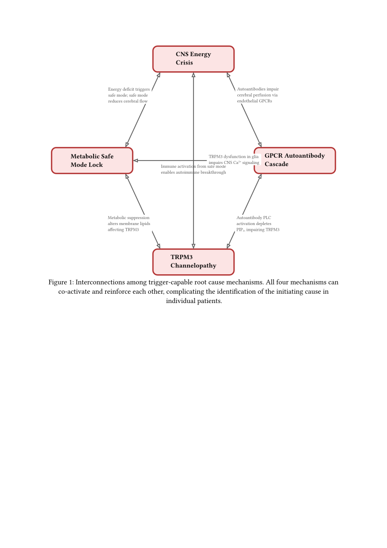
The four trigger-capable mechanisms are not independent. Their interconnections are extensive and create a web of potential co-activation that complicates the question of “which came first” while simultaneously explaining why ME/CFS is so resistant to single-target treatments.
GPCR autoantibodies may modulate TRPM3 gating through second messenger pathways: muscarinic receptor autoantibodies activate phospholipase C, which hydrolyzes PIP2—a lipid required for TRPM3 channel activity (Section Novel Hypotheses from TRPM3 Ion Channel Research). Chronic PIP2 depletion from sustained autoantibody-driven PLC activation could reduce TRPM3 function, making the GPCR autoantibody cascade and TRPM3 channelopathy two aspects of a single pathogenic process rather than independent root causes.
CNS energy failure may activate the metabolic safe mode program: if hypothalamic energy supply drops below a threshold, the brain may interpret this as a threat signal and engage the metabolic suppression program, linking the CNS energy crisis to the safe mode lock. Conversely, the safe mode program’s deliberate metabolic suppression includes reduced cerebral blood flow and neurotransmitter synthesis, contributing to the CNS energy deficit.
The safe mode program’s immune activation component—maintained cytokine production, persistent inflammatory signaling—creates an environment conducive to autoimmune breakthrough through molecular mimicry or bystander activation, potentially generating GPCR autoantibodies as a secondary consequence of the safe mode state.
These interconnections mean that in practice, trigger-capable mechanisms may co-activate, and the question of which mechanism was “first” may be unanswerable in individual patients. The more productive question—and the one with therapeutic relevance—is: which root-level processes are currently active and maintaining the disease? A patient whose disease began with GPCR autoantibodies may now be maintained primarily by the metabolic safe mode lock, if epigenetic consolidation has rendered the safe mode self-sustaining independent of its original autoantibody trigger. Conversely, a patient who entered through the safe mode door may now have developed secondary GPCR autoantibodies that represent a new, independent disease-maintaining mechanism.
This distinction between initiating cause and maintaining cause is crucial for treatment strategy and is developed further in Section Treatment Implications of the Causal Hierarchy.
Figure trigger mechanism relationships maps the bidirectional interactions among the four trigger-capable mechanisms, illustrating how co-activation and mutual reinforcement complicate the identification of a single initiating cause.