Central Nervous System Abnormalities
1 Brain Structure and Function
1.1 Structural Neuroimaging Findings
Multiple neuroimaging studies have documented structural brain abnormalities in ME/CFS patients, though findings have varied across studies due to differences in patient populations, imaging protocols, and analytical methods (J. Lee et al. 2024).
White Matter Abnormalities Several studies have reported increased white matter hyperintensities (WMH) in ME/CFS patients compared to healthy controls (Lange et al. 1999) (Zeineh et al. 2015). These hyperintensities, visible on T2-weighted and FLAIR MRI sequences, may indicate demyelination, axonal loss, or microvascular damage. The distribution of WMH in ME/CFS patients tends to involve periventricular white matter, subcortical regions, and frontal and temporal lobes. Zeineh et al. (Zeineh et al. 2015) identified increased fractional anisotropy in the right arcuate fasciculus, which correlated with disease severity (r=0.649, p=0.0015), providing anatomical substrate for the cognitive dysfunction observed in ME/CFS.
The clinical significance of these findings remains debated, as similar changes occur with normal aging and various medical conditions. However, the presence of WMH in younger ME/CFS patients suggests pathological processes beyond typical age-related changes (Lange et al. 1999) (Zeineh et al. 2015).
1.2 Diffusion-Based White Matter Abnormality Imaging
Yu et al. (2026) applied an advanced neuroinflammation imaging (NII) model to diffusion MRI data from 67 ME/CFS patients and 67 matched healthy controls (Yu2026diffusion?)-neuroinflammation. The NII model estimates tissue microstructure parameters beyond conventional diffusion tensor imaging (DTI) and revealed three abnormalities: reduced NII-HR (consistent with cerebral edema), reduced NII-RF (consistent with cellular infiltration), and increased NII-FF (consistent with axonal reorganisation). These parameters cannot definitively distinguish neuroinflammation from other white matter pathologies—chronic cerebral hypoperfusion (documented in 90% of ME/CFS patients, Section Cerebral Blood Flow Abnormalities) could produce the same pattern through ischemia-reperfusion injury without any immune mechanism.
Certainty: 0.35. n=67 per group with rigorous matching; NII model substantially more sensitive than conventional DTI; findings correlate with clinical measures; Human Brain Mapping is a reputable specialty journal. Not independently replicated. Certainty at 0.35 (not 0.45) because: single-center cross-sectional study; NII model has not been validated in independent ME/CFS cohorts or against histopathology in any disease; the “neuroinflammation” interpretation is one of several plausible explanations (ischemia, demyelination, neurodegeneration could produce identical NII signatures); conventional DTI showing minimal differences could indicate NII is oversensitive rather than that DTI missed real pathology; hypoperfusion already well-documented in ME/CFS provides an equally parsimonious explanation without any novel model. (Yu2026diffusion?)-neuroinflammation
The critical methodological advance is that conventional DTI metrics showed minimal group differences, while NII metrics revealed widespread abnormalities. This resolves a longstanding inconsistency in ME/CFS neuroimaging—prior DTI studies produced mixed results, which may have reflected insufficient sensitivity of the standard DTI model rather than absence of pathology. The NII model’s parameters map more directly onto tissue-level processes (edema, cellular infiltration, axonal change), providing a clearer window into the biological substrate of white matter abnormalities.
The NII findings are broadly consistent with neuroinflammation, chronic hypoperfusion, or a combination. The cerebral hypoperfusion extensively documented in ME/CFS (Section Cerebral Blood Flow Abnormalities: 10–20% global CBF reduction, 90% of patients with abnormal tilt-test CBF) provides a parsimonious alternative explanation: ischemia-reperfusion injury from episodic hypoperfusion could produce the edema, cellular infiltration, and axonal reorganisation patterns observed without requiring an immune mechanism. Distinguishing neuroinflammation from ischemia will require either histopathological correlation or multi-modal imaging (e.g., TSPO-PET combined with NII).
NII analysis is a research technique performed with specialised post-processing software; it is not available through clinical radiology departments, and standard diffusion MRI cannot substitute.
Limitations: Cross-sectional design precludes causal inference; single-center study; NII model requires validation in independent ME/CFS cohorts; the neuroinflammation interpretation, while plausible, is inferential rather than directly confirmed by histopathology; hypoperfusion (already well-documented in ME/CFS) is an equally parsimonious explanation for the observed NII parameters.
Certainty: N/A — the direction of any brainstem volume difference is unresolved. A proof-of-concept multimodal MRI study (Vienna, n=26 ME/CFS / 27 controls) reported reduced brainstem volume including the pons in patients: \(\beta\)=-0.10% eTIV (95% CI -0.18 to -0.02, \(p\)=0.013, FDR-\(p\)=0.039) (Bader et al. 2026). Deep grey matter and whole-brain parenchymal fraction did not differ (FDR 0.98), indicating a brainstem-specific, not global, atrophic signal. By contrast, an independent 7T MRI study from the Griffith University lab reported larger brainstem volumes in ME/CFS and long COVID (pons \(p\)=0.003, superior cerebellar peduncle \(p\)=0.009, whole brainstem \(p\)=0.005) (Thapaliya et al. 2023). These two findings point in opposite directions.
The contradiction is not resolved by study quality alone: both use structural MRI in ME/CFS cohorts. Per the integration decision for this topic, the direction is presented as an open question rather than weighted toward either study. The cohort-overlap caveat matters: Thapaliya2023, Barnden2018, and Thapaliya2022 originate from the same Griffith laboratory cluster, so they are not independent replications of each other (Thapaliya et al. 2023) (Barnden et al. 2018) (Thapaliya et al. 2022). Accounting for this, the net independent clusters number two (Vienna reduced vs. Griffith larger) with opposite directions.
Replication status: Each direction rests on a single independent research group; not reconciled across independent labs.
Severity applicability: Unknown — neither cohort stratified brainstem volume by disease severity; any relationship between brainstem volume direction and symptom burden is untested.
Limitations: Proof-of-concept and modest sample in the Vienna study; possible methodological differences (field strength 3T vs 7T, segmentation pipelines, cohort composition); no study yet measures brainstem volume and function (e.g., connectivity, metabolite levels) in the same patients to reconcile direction with symptom burden.
Falsifiable prediction: A multi-site study using harmonised segmentation across an adequate sample will determine whether brainstem volume is reduced, increased, or unchanged in ME/CFS, and whether any direction correlates with severity, disease duration, or brainstem-specific symptoms (autonomic dysfunction, dyspnoea, sleep disturbance). The open question is resolved once independent labs using comparable methods obtain a consistent direction.
Consequence: The two groups’ opposite results mean we cannot yet trust any single claim about brainstem size in ME/CFS — knowing the true direction matters because a consistent structural brainstem finding would point to a specific site of pathology and help reconcile the many brainstem-related symptoms patients report.
Certainty: N/A — the biological substrate of the elevated T1w/T2w ratio signal is unresolved. A multimodal MRI study from the Griffith University lab compared long COVID (n=19), COVID-recovered healthy controls (n=12), and healthy controls without COVID (n=16), finding significantly altered T1w/T2w ratio signal (a proxy for myelin content) between all three groups across several regions: long COVID showed higher signal than controls in the precentral gyrus and middle temporal gyrus, recovered controls showed higher signal than long COVID in the pons, midbrain, cerebellar tonsil, superior longitudinal fasciculi, and precentral gyrus, and recovered controls showed higher signal than never-infected controls in the precentral gyrus and posterior cingulate cortex (Thapaliya et al. 2025). The T1w/T2w signal in the middle temporal gyrus correlated with physical function (\(r = 0.56\)) and in the midbrain with cognitive score (\(r = -0.64\)) (Thapaliya et al. 2025).
The authors interpret the elevated signal as possible remyelination, but explicitly concede it “may also indicate inflammation or gliosis” (Thapaliya et al. 2025). This is the same interpretive ambiguity that limits all T1w/T2w-based myelin claims: the ratio is sensitive to iron content, water, and tissue structure, not myelin alone, so elevated signal cannot distinguish remyelination from neuroinflammatory gliosis or iron deposition. The finding echoes the same group’s earlier ME/CFS report of elevated T1w/T2w in white matter and subcortical grey matter (Thapaliya et al. 2020), indicating a consistent lab-level signal but not yet an independent or mechanistically resolved one.
Replication status: The elevated-T1w/T2w finding rests on a single research group (the Griffith NCNED cluster: Thapaliya 2025, Thapaliya 2020); the three-group design (long COVID vs recovered vs never-infected) is novel, but no independent lab has replicated the specific regional pattern.
Competing explanations: (1) Remyelination — increased myelin after viral injury; (2) neuroinflammatory gliosis — reactive glia increasing signal independent of myelin; (3) iron deposition or water-content changes — non-myelin contributors to the T1w/T2w ratio. The study cannot separate these without a myelin-specific or iron-specific sequence. The three-group direction is itself informative: the recovered group (not the symptomatic long-COVID group) showed the highest signal in the pons, midbrain, cerebellar tonsil, and superior longitudinal fasciculi — which, if elevated signal tracks myelin, would point to remyelination/recovery rather than damage (long COVID’s relatively lower brainstem signal then reflecting failed or delayed remyelination). Iron is the strongest single alternative: T1w/T2w is directly iron-sensitive (tissue iron shortens T2 and raises the ratio, mimicking a myelin reading), and post-COVID iron dyshomeostasis is independently documented (persistent hyperferritinaemia and iron-handling abnormalities up to a year after infection (Sonnweber et al. 2022) (Kronstein-Wiedemann et al. 2024)) — so a metal-handling explanation is more than a tie. Under the iron reading, the “remyelination/recovery” interpretation collapses and the signal becomes a marker of iron/metabolic handling, not repair.
Severity applicability: Unknown — none of the cohorts was stratified by disease severity; the long-COVID group was not phenotyped for ME/CFS diagnostic criteria.
Limitations: Cross-sectional; small subgroups (n=12–19); exploratory cluster-based analysis with possible Type I error inflation; single lab (the Griffith NCNED cluster — Thapaliya 2025, 2020 and related DTI papers are not independent replications of each other); no longitudinal follow-up; no myelin water imaging, quantitative susceptibility mapping, or astrogliosis-specific PET to disambiguate the substrate. The structure–function correlations rest on just two correlations (r = 0.56, r = −0.64) in n = 47 with no diffusion–clinical correlation surviving correction, so they should be treated as preliminary, not established. The COVID-recovered group are a selected resilient subset (survivors without persistent symptoms), and the cohort is WHO-long-COVID, not ME/CFS-diagnosed — so no finding transfers to ME/CFS without a direct ME/CFS replication.
Falsifiable prediction: A study combining T1w/T2w with myelin water fraction, quantitative susceptibility mapping, and an astrogliosis-specific readout (e.g., the astroglial MAO-B PET tracer [11C]SL25.1188; TSPO-PET alone is an inadequate gliosis probe because long-COVID TSPO is heterogeneous and regionally specific (Braga et al. 2025) (Visser et al. 2025)) in the same long-COVID / recovered cohort will determine whether the elevated signal tracks myelin content (remyelination), gliosis, or iron — resolving whether the finding reflects recovery or ongoing pathology. A longitudinal rescan of the same cohort (rather than cross-sectional comparison of different groups) is the decisive test: it separates “recovery causes remyelination” from “recoverers were constitutionally different from the outset.” The open question is resolved once an independent lab confirms the regional pattern and a myelin/iron-specific method identifies the substrate.
Consequence: Whether the elevated brain-signal in long COVID and recovered patients reflects healing (remyelination) or ongoing damage (inflammation or scarring) changes what the scan means — if it is inflammation or gliosis, it points to persistent brain pathology that may warrant monitoring, whereas if it is remyelination it suggests recovery.
2 CSF Proteomics: Complement, Coagulation, and Clinical Heterogeneity
Bragee et al. (2026) performed CSF proteomics on 31 ME/CFS patients, quantifying 902 proteins and correlating findings with clinical features Bragée et al. (2026). The most striking finding was the enrichment of neutrophil degranulation and platelet activation pathways in POTS-positive patients, alongside complement cascade and coagulation pathway enrichment in severe cases.
This CSF compartment data is valuable because it samples the central nervous system directly. However, the absence of a control group limits interpretation: the pathway enrichments (neutrophil degranulation, platelet activation, complement cascade, coagulation) are associated with clinical features within the ME/CFS group but cannot be established as different from healthy individuals. Furthermore, complement and coagulation proteins are among the most abundant plasma proteins, and blood contamination is a dominant confound in CSF proteomics—if BBB permeability is increased in ME/CFS (as documented in Section Spike Protein Primes Brain Innate Immunity, Lowering the Neuroinflammatory Threshold), plasma protein leakage would be expected and does not necessarily indicate CNS-compartment-specific pathology. Within-group severity correlations are directionally consistent with peripheral blood findings but cannot establish cross-compartment convergence without a control group.
Certainty: 0.35. n=31 ME/CFS patients with no healthy control group; ratio-based within-group analysis only; single-center; Scientific Reports is mid-tier. The absence of a control group means differential expression between patients and healthy individuals cannot be established; only within-patient severity associations are possible. These findings require validation in an independent cohort with matched controls.
Gray Matter Volume Changes Voxel-based morphometry (VBM) studies have identified regional brain abnormalities in ME/CFS patients, though findings vary across cohorts (Puri et al. 2012) (Finkelmeyer et al. 2017) (Shan et al. 2020). Documented changes include gray matter differences in the parahippocampal gyrus, occipital regions, amygdala, and insula, alongside white matter volume reductions in the brainstem and temporal regions. No single pattern has been consistently replicated across studies, reflecting the clinical heterogeneity of ME/CFS.
Despite this heterogeneity, the presence of structural brain differences in regions involved in memory, interoception, and autonomic regulation supports a neuroanatomical basis for cognitive and autonomic dysfunction in ME/CFS (Shan et al. 2020).
2.1 Functional Neuroimaging: The NIH Deep Phenotyping Study
The 2024 NIH study by Walitt et al. employed functional MRI during motor tasks to identify specific brain regions with abnormal activation patterns in PI-ME/CFS patients (Walitt, Singh, LaMunion, Hallett, et al. 2024). This study, involving 17 PI-ME/CFS patients and 21 matched healthy controls, provided the most rigorous functional neuroimaging data to date.
Temporal-Parietal Junction Dysfunction
Walitt et al. (Walitt, Singh, LaMunion, Hallett, et al. 2024) identified abnormally reduced activity in the temporal-parietal junction (TPJ) during effort-based decision-making tasks in PI-ME/CFS patients. The TPJ is a heteromodal association cortex that integrates information from multiple sensory modalities and plays essential roles in agency and intention attribution, effort allocation decisions, attentional reorienting, social cognition, and bodily self-consciousness. This dysfunction provides a neuroanatomical substrate for the characteristic mismatch between perceived capability and actual performance that defines ME/CFS, suggesting the brain genuinely perceives effort requirements inaccurately rather than exhibiting malingering or simple deconditioning.
Single study (Walitt 2024, n=17 PI-ME/CFS vs. 21 controls). This finding derives from the NIH deep phenotyping study—the first to examine TPJ function in ME/CFS. The small sample size limits generalizability. Independent replication in a larger cohort and across ME/CFS subtypes is required before TPJ dysfunction can be considered an established feature.
Motor Cortex Hyperactivity Paradoxically, while the TPJ showed reduced activation, the motor cortex demonstrated sustained hyperactivity during fatiguing grip tasks in ME/CFS patients (Walitt, Singh, LaMunion, Hallett, et al. 2024). The motor cortex remained abnormally active despite declining grip force output, yet electromyography showed no evidence of peripheral muscle fatigue. This dissociation between central motor drive and peripheral performance reveals inefficient neural recruitment patterns requiring excessive cortical activation for submaximal force production.
This pattern indicates that fatigue in ME/CFS originates centrally rather than peripherally (Walitt, Singh, LaMunion, Hallett, et al. 2024). The motor cortex continues to “try harder” even as actual force production declines, suggesting a breakdown in the feedback mechanisms that normally calibrate effort to output. A complementary multimodal study (Bedard 2026) found that ME/CFS patients fail to up-regulate motor output (cortical and subcortical) under sustained effort despite equal maximum force (Bedard et al. 2026); the resulting central motor-drive failure and its differential-diagnostic drug probes are traced in Section Systemic inflammation is downstream of a more upstream cause.
Effort Preference Alteration: A New Paradigm Perhaps the most conceptually important finding from the NIH study was the identification of altered effort preference as a defining feature of PI-ME/CFS, distinct from physical fatigue (muscle exhaustion) or central fatigue (reduced motor cortex output). Walitt et al. proposed that:
“Fatigue may arise from a mismatch between what someone thinks they can achieve and what their bodies perform.”
This reconceptualization has profound implications for understanding ME/CFS. First, the brain genuinely perceives effort requirements inaccurately, leading to appropriate behavioral responses to faulty signals; this rules out malingering or simple deconditioning. Second, the TPJ normally synthesizes multiple information streams—interoceptive, proprioceptive, and motivational—to generate effort estimates, but this integration fails in ME/CFS. Third, the brain may be responding to genuine danger signals such as inflammation or metabolic dysfunction while miscalibrating the protective response. Finally, interventions targeting effort perception and decision-making networks may prove more effective than those addressing peripheral fatigue.
Certainty: 0.50. ME/CFS symptoms may represent an evolutionarily conserved “sickness behavior” program—normally protective during acute infection—that becomes chronically activated due to persistent immune signaling. The TPJ, which normally integrates inflammatory signals with effort allocation decisions, may misinterpret chronic low-grade inflammation as ongoing acute illness, inappropriately suppressing activity to “conserve resources” for an immune battle that has already concluded (or that persists at subclinical levels). This would explain why the fatigue feels so viscerally “real” and protective to patients: the brain is executing a legitimate survival program, but one triggered by faulty or persistent signals rather than current metabolic necessity.
Risk-Based Decision-Making Impairment During behavioral tasks requiring risk assessment and effort allocation, ME/CFS patients demonstrated reduced selection of “hard” task options even when rewards were equivalent, difficulty sustaining effort on extended tasks, and altered subjective perception of task difficulty (Walitt, Singh, LaMunion, Hallett, et al. 2024). Notably, motivation levels remained normal despite reduced effort output.
These findings indicate that the problem lies not in willingness to exert effort (motivation) but in the neural computation of what constitutes acceptable effort levels (Walitt, Singh, LaMunion, Hallett, et al. 2024).
2.2 PET Scan Metabolic Findings
Positron emission tomography (PET) studies have revealed regional hypometabolism in ME/CFS patients, indicating reduced glucose utilization and neuronal activity (Chaudhuri and Behan 2004). Commonly affected regions include brainstem nuclei (potentially explaining autonomic dysfunction), basal ganglia (correlating with motor symptoms and fatigue), medial prefrontal cortex (associated with executive dysfunction), and posterior parietal cortex (linked to attention and spatial processing deficits).
The pattern of hypometabolism overlaps significantly with regions showing structural and functional abnormalities, consistent with a coherent picture of multifocal brain dysfunction.
Correlation of imaging findings does not establish that these brain abnormalities cause ME/CFS symptoms, nor which abnormality precedes others. Temporal precedence, reverse causation (systemic illness causing brain changes), and potential confounders (medication effects, deconditioning, sleep disruption) require further investigation through longitudinal and interventional neuroimaging studies.
2.3 SPECT Perfusion Abnormalities
Single-photon emission computed tomography (SPECT) studies have documented reduced regional cerebral blood flow (rCBF) in ME/CFS patients C. (Linda). M. C. van Campen, Rowe, and Visser (2020). Characteristic findings include global reduction in cortical perfusion (10–15% below controls), focal hypoperfusion in temporal, frontal, and parietal regions, correlation between perfusion deficits and cognitive symptom severity, and exacerbation of perfusion abnormalities following physical or cognitive exertion.
The persistence of perfusion deficits across multiple studies and imaging modalities is consistent with cerebrovascular dysfunction contributing to ME/CFS symptoms. Cataldo et al. (2026, n=186) confirm this extends to Long COVID: ASL MRI revealed increased spatial coefficient of variation (sCOV) indicating delayed arterial transit and global microvascular dysfunction — despite preserved cerebral blood flow, suggesting the pathology is microvascular efficiency rather than gross perfusion deficit (Cataldo et al. 2026).
The CSF catecholamine data derive primarily from two NIH Clinical Center analyses of overlapping cohorts: Walitt 2024 (n=17 PI-ME/CFS) and Aregawi 2026 (n=16 PI-ME/CFS, n=34 PASC, n=32 Parkinson’s disease, n=40 healthy volunteers) (Walitt, Singh, LaMunion, and others 2024) (Aregawi et al. 2026). Aregawi et al. extended the 2024 findings by constructing novel composite pathway indices (NE + DHPG + MHPG for noradrenergic; DA + DOPAC + HVA for dopaminergic), adding PASC and Parkinson’s disease comparator groups, and reporting CSF MHPG levels for the first time in PI-ME/CFS or PASC. Key epistemic boundaries:
- Both studies originate from the same NIH Clinical Center; no independent replication at a different institution has been performed.
- The cohorts were restricted to post-infectious ME/CFS; whether noradrenergic deficiency characterises gradual-onset or non-viral ME/CFS is unknown.
- CSF catecholamine levels are influenced by medications, sleep timing, activity levels, and lumbar puncture technique — though Aregawi et al. demonstrated the NE Pathway abnormality persisted after removing patients on tricyclics, amphetamines, venlafaxine, or duloxetine.
- The direction of causality is unestablished: low central norepinephrine may result from reduced physical activity, sleep disruption, or chronic illness rather than causing ME/CFS symptoms. Prolonged inactivity reduces CSF NE turnover in healthy individuals, and ME/CFS patients are among the most sedentary clinical populations. Neither the Walitt 2024 nor Aregawi 2026 cohorts included deconditioning-matched controls, a confound that has not been experimentally eliminated. The correlation pattern is consistent with this alternative: NE Pathway correlates with handgrip endurance (a readout of physical conditioning, rho=0.62) but not with orthostatic measures (the symptom domain most directly linked to NE physiology), a dissociation expected if low CSF NE tracks deconditioning rather than driving pathophysiology.
- Adrenergic dysfunction is broadly supported: a systematic review and meta-analysis by Hendrix et al. (2025) confirmed adrenergic dysfunction across ME/CFS and fibromyalgia, though with heterogeneous measures and outcomes (Hendrix et al. 2025).
- The Hendrix 2025 meta-analysis reports elevated catecholamines and altered receptor function — the opposite pattern from the low CSF NE Pathway reported by Aregawi 2026. This tension (elevated peripheral NE vs. reduced central NE) is consistent with the central-peripheral mismatch model but could also reflect different populations, measurement timing, or NE redistribution rather than production failure.
3 Brain as Energy Coordination Bottleneck
The convergence of regional hypometabolism (PET), hypoperfusion (SPECT), neuroinflammation, and catecholamine deficiency raises a fundamental question: is brain dysfunction in ME/CFS secondary to systemic illness, or could it be primary—the bottleneck limiting whole-body function?
Certainty: 0.45. The near-universal presence of cognitive dysfunction, documented brain hypometabolism (Chaudhuri and Behan 2004), and neuroinflammation with 45–199% elevation in key regions (Nakatomi et al. 2014) suggest CNS energy crisis may be the primary pathophysiological event. Direct in-vivo measurement now adds a molecular energy readout: reduced ATP-to-phosphocreatine ratios on ³¹P-MRSI in Long COVID (see Clinical Finding In Vivo CNS ATP and Phosphocreatine Reduction in Long COVID) provide among the first direct evidence of a measurable brain high-energy-phosphate deficit in post-infectious fatigue (Weber-Fahr et al. 2026). The Maccallini 2026 meta-GWAS provides independent genetic support for this model: ME/CFS genetic risk variants are enriched exclusively in brain and pituitary tissues across 30 tissue types, with no peripheral tissue reaching significance (Maccallini 2026). Failure of the brain to coordinate peripheral demand-responsive processes could explain the selective pattern of dysfunction observed in ME/CFS, where autonomous processes (hair growth, nail growth, baseline cellular metabolism) remain intact while CNS-coordinated responses (exercise capacity, orthostatic tolerance, immune adaptation) are severely impaired.
Testable prediction: CNS-targeted interventions (microglial modulation, catecholamine restoration, cerebral perfusion enhancement) should outperform periphery-only interventions (mitochondrial cofactors without CNS penetration, peripheral immune modulation) on global symptom burden in randomised comparative trials. Falsified if periphery-targeted treatments produce equivalent or greater whole-body improvement without CNS-specific effects. See Neuroinflammatory Cascade: From CNS to Peripheral Symptoms for the detailed causal cascade prediction.
Weber-Fahr et al. (2026) used whole-brain phosphorus magnetic resonance spectroscopy (31P-MRSI) to measure high-energy phosphates directly in living patients, comparing 27 post-COVID participants with 23 recovered controls (Weber-Fahr et al. 2026). Long COVID participants showed reduced ATP-to-phosphocreatine (ATP/PCr) ratios centred on the cingulate cortex, with lower anterior-cingulate ATP/PCr correlating with poorer cognition. The Long COVID subgroup meeting ME/CFS diagnostic criteria showed the same brain-energy alteration. This is among the first direct in-vivo human evidence that brain ATP and its phosphagen reserve are measurably reduced in post-infectious fatigue—complementing the PET hypometabolism and neuroinflammation findings above with a molecular energy readout rather than an indirect perfusion or metabolic proxy. (Evidence type: observational case-control; raw certainty 0.72, from a Long-COVID cohort with an ME/CFS subgroup — discounted to 0.61 for the cross-cohort transfer. Correlational, not causal.) (Severity applicability: unknown — the study did not stratify by ME/CFS severity.)
Reading this as a core ME/CFS energy lesion is premature: the deficit may be specific to the Long-COVID trajectory rather than general ME/CFS, could be a downstream correlate of reduced cognitive activity or depressive/medication state rather than an upstream energy-generating failure, and the ATP/PCr-cognition link is correlational and potentially reverse-causal (Weber-Fahr et al. 2026). Until it replicates in non-COVID ME/CFS and survives control for cognitive effort and mood, it is best treated as a candidate biomarker of the central-fatigue phenotype, not proof of a primary brain energy-generating defect.
Consequence: If confirmed in broader ME/CFS cohorts, direct in-vivo brain-energy measurement would give researchers a molecularly specific readout of the CNS energy deficit this model predicts — currently inferred only from perfusion and metabolism proxies.
3.1 Evidence for Brain-Centric Model
Several observations support the brain as primary bottleneck:
Universal cognitive involvement: Brain fog and cognitive dysfunction are present in nearly all ME/CFS patients, regardless of primary symptom presentation or disease severity (Walitt, Singh, LaMunion, Hallett, et al. 2024)
Disproportionate brain energy demand: The brain comprises 2% of body mass but consumes 20–25% of resting energy (Magistretti and Allaman 2018), making it uniquely vulnerable to energy constraint
Cascading coordination failure: The brain coordinates peripheral demand responses via autonomic signaling; CNS energy deficit would impair this coordination across multiple organ systems simultaneously
Catecholamine deficiency: Reduced CSF catecholamines (Section neurotransmitters) directly impair the signaling required for demand-response mobilization throughout the body
Neuroinflammation evidence: Nakatomi et al. (Nakatomi et al. 2014) documented 45–199% elevation in neuroinflammatory markers across six brain regions (cingulate cortex, hippocampus, amygdala, thalamus, midbrain, pons), with inflammation severity correlating directly with cognitive impairment and pain. While replication of these PET findings remains incomplete (see Section Sleep EEG Delta/Alpha Ratio as a Non-Invasive Thalamic Calcium Proxy), the magnitude and regional distribution suggest potential CNS-specific pathology warranting further investigation
3.2 Autonomic Dysfunction as Downstream Effect
If the brain cannot maintain adequate energy for autonomic coordination, peripheral organs would have energy available but lack the signals to mobilize it appropriately. This explains several key observations:
Pharmacological bypass efficacy: Midodrine, fludrocortisone, and other autonomic-supporting medications can partially restore function—the peripheral targets respond when appropriately stimulated, suggesting the dysfunction is in coordination rather than peripheral capacity
Demand-response failure pattern: Baseline function often preserved while challenge responses fail; the CNS cannot orchestrate the coordinated scaling required for physiological stress
Preservation of autonomous processes: Truly local processes (hair follicle cycling, which operates an independent internal Cori cycle) continue unaffected because they don’t require CNS coordination
3.3 Cerebral Blood Flow: The Central Vulnerability
Van Campen and colleagues have systematically documented cerebral blood flow (CBF) abnormalities during orthostatic stress that support the brain-centric model (C. L. M. C. van Campen et al. 2020) (C. L. M. C. van Campen, Rowe, and Visser 2021) (C. L. M. C. van Campen, Rowe, and Visser 2023) (C. L. M. C. van Campen et al. 2024):
In a series of studies using transcranial Doppler during tilt-table testing, van Campen et al. demonstrated that 91% of ME/CFS patients (488/534) with normal heart rate and blood pressure responses show abnormal cerebral blood flow and cardiac output reduction during orthostatic challenge (C. L. M. C. van Campen et al. 2024). The magnitude of CBF decline is approximately 3.7-fold greater than healthy controls (26% vs. 7% reduction at end-tilt) (C. L. M. C. van Campen et al. 2020). Furthermore, CBF remains reduced even after returning to supine position, with recovery correlating to disease severity rather than hemodynamic parameters (C. L. M. C. van Campen, Rowe, and Visser 2021). For detailed breakdown by orthostatic phenotype, see Achievement Near-Universal CBF Decline in ME/CFS in Chapter Cardiovascular Dysfunction.
All CBF findings derive from a sequential publication series by van Campen and colleagues at a single Dutch referral center (4 papers, 2020–2024). While the large cumulative sample (n=534) and consistent methodology strengthen internal validity, no independent research group has replicated these transcranial Doppler findings during tilt-table testing. The appearance of replication across multiple publications reflects one group’s expanding cohort, not independent confirmation. Independent replication at other centers with different equipment and patient populations is essential.
ME/CFS symptom severity correlates directly with the degree of CBF reduction during tilt testing (C. L. M. C. van Campen, Rowe, and Visser 2023). Patients with greater CBF decline report worse fatigue, cognitive dysfunction, and orthostatic symptoms. The absence of compensatory cerebral vasodilation despite reduced cardiac output suggests possible endothelial dysfunction contributing to cerebrovascular vulnerability (C. L. M. C. van Campen et al. 2024) (Medow and Stewart 2024) (Badhwar et al. 2025). The brain’s high metabolic demand and sensitivity to perfusion deficits may make cerebral blood flow the “canary in the coal mine” for systemic energy coordination dysfunction. Standard vital sign monitoring during orthostatic challenge misses this pathology—normal heart rate and blood pressure do not exclude significant cerebrovascular compromise.
See Chapter Energy Metabolism and Mitochondrial Function Section Selective Energy Dysfunction: The CNS-Dependency Hypothesis for integration with the selective energy dysfunction hypothesis, and Chapter Cardiovascular Dysfunction Section Near-Universal Vasopressin Deficiency in ME/CFS for detailed cerebrovascular findings.
4 Neurotransmitter Abnormalities
The structural and functional brain abnormalities described above correlate with specific neurochemical deficits identified in the NIH deep phenotyping study, providing mechanistic links between imaging findings and clinical symptoms.
4.1 Catecholamine Pathway Dysregulation: CSF Findings
The NIH deep phenotyping study provided the first direct evidence linking cerebrospinal fluid (CSF) catecholamine abnormalities to ME/CFS symptoms (Walitt, Singh, LaMunion, Hallett, et al. 2024). This represents a major advance in understanding the neurochemical basis of the disease.
Reduced CSF Catecholamines
The 2024 NIH deep phenotyping study provided the first direct evidence from CSF analysis linking central catecholamine abnormalities to ME/CFS symptoms, documenting reduced CSF DOPA, DOPAC, and DHPG (Walitt, Singh, LaMunion, and others 2024). A follow-up analysis by Aregawi et al. (2026) refined these findings with a novel composite pathway-index methodology, constructing Norepinephrine Pathway (NE + DHPG + MHPG) and Dopamine Pathway (DA + DOPAC + HVA) indices applied across four groups: PI-ME/CFS (n=16), PASC/Long COVID (n=34), Parkinson’s disease (n=32), and healthy volunteers (n=40) (Aregawi et al. 2026).
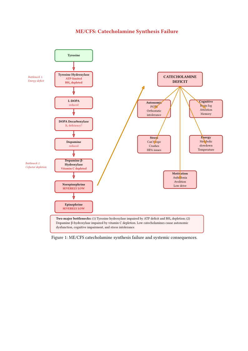
The key advance is the demonstration of selective noradrenergic deficiency. Mean Norepinephrine Pathway activity was significantly lower in PI-ME/CFS and PASC with post-exertional malaise compared to healthy volunteers, while the Dopamine Pathway index showed no significant difference between patient groups and controls. This selectivity argues against a global catecholamine synthesis failure (e.g., BH4 depletion affecting both pathways equally) and points instead to a noradrenergic-specific defect, consistent with the ATP-dependent conversion of dopamine to norepinephrine within vesicles by dopamine-beta-hydroxylase.
CSF MHPG, the main end-product of central norepinephrine metabolism and the predominant analyte in the NE Pathway index, was significantly lower in PI-ME/CFS and PASC compared to controls — the first report of CSF MHPG levels in either condition. The authors speculate that decreased ATP availability impairs the vesicular proton pump required for dopamine-beta-hydroxylase activity, selectively blocking the conversion of dopamine to norepinephrine while leaving cytoplasmic dopamine metabolism intact.
The DHPG finding is particularly significant because it is the primary intraneuronal metabolite of norepinephrine produced within noradrenergic neurons. Low CSF DHPG specifically indicates reduced norepinephrine turnover in the central nervous system, pointing to hypofunction of the locus coeruleus and other noradrenergic nuclei. The selective NE-over-DA pathway deficiency is consistent with preserved dopaminergic synthesis in PI-ME/CFS, though new VMAT2 PET evidence in long COVID (Observation Striatal Dopaminergic Terminal Loss Quantified by VMAT2 PET in Long COVID) raises the possibility of dopaminergic terminal loss that may not be captured by CSF metabolite indices alone — resolving this tension requires direct VMAT2 PET in ME/CFS.
Partially replicated (same NIH Clinical Center, extended with novel composite pathway index). Aregawi et al. (2026) confirmed the noradrenergic deficiency using a refined methodology (composite NE + DHPG + MHPG index) and extended findings to PASC/Long COVID patients (Aregawi et al. 2026). The selectivity for noradrenergic over dopaminergic pathways is consistently demonstrated across both analyses. However, no independent replication at a different institution using a distinct patient cohort has been performed; the invasiveness of CSF collection limits feasibility of large-scale multi-center replication. The fundamental question of temporal stability — whether CSF catecholamine deficiency is stable trait or fluctuates with disease phase — also remains unaddressed.
Clinical Correlations
Walitt et al. (Walitt, Singh, LaMunion, Hallett, et al. 2024) established direct correlations between CSF catecholamine levels and clinical measures. Lower catecholamines correlated with reduced grip strength endurance and slower reaction times (motor performance), catecholamine deficits predicted reduced selection of hard tasks in decision-making paradigms (effort-related behaviors), memory and executive function scores correlated with dopamine metabolite levels (cognitive impairment), and subjective fatigue ratings inversely correlated with norepinephrine concentrations (fatigue severity).
Aregawi et al. (2026) extended these correlations using the composite NE Pathway index (Aregawi et al. 2026). Across all participants (PI-ME/CFS, PASC, and healthy volunteers, n=63), NE Pathway activity correlated positively with handgrip duration (right hand: rho=0.54, P=4e-5; left hand: rho=0.40, P=0.0027), PROMIS mental and physical health scores, and SF-36 physical functioning, vitality, and general health sub-scores. Significant negative correlations were found with PROMIS fatigue, MFI general and physical fatigue sub-scores, and emotional role limitations. Within the patient-only analysis (n=38), the strongest associations were handgrip duration (right: rho=0.62, P=0.00011) and MFI physical fatigue (rho=-0.43, P=0.0094). Notably, NE Pathway activity did not correlate with pain, cognition measures, anxiety, or depression scores, and showed no relationship with orthostatic hypotension or excessive orthostatic tachycardia, suggesting the deficiency is more closely tied to motor output and overall health perception than to autonomic cardiovascular regulation per se.
These correlations suggest, for the first time in direct CSF measurement, a biochemical pathway linking specific neurotransmitter abnormalities to the core symptoms of ME/CFS.
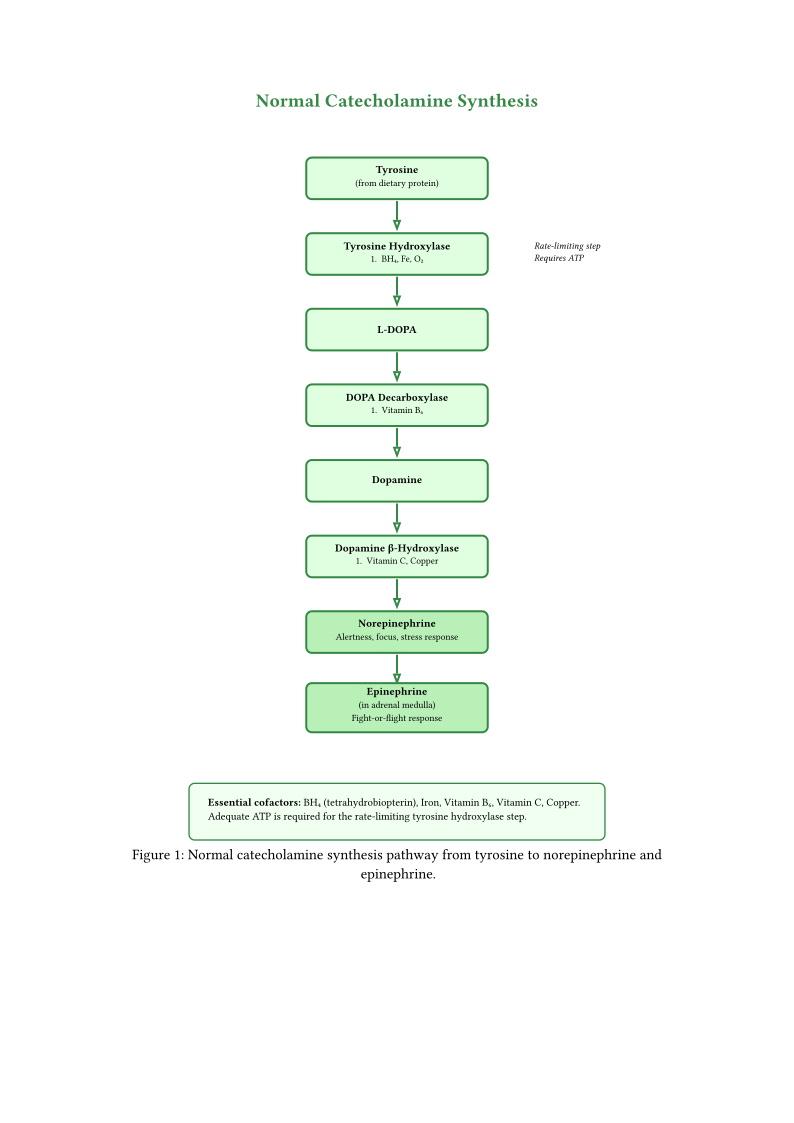
Figures catecholamine normal and catecholamine mecfs illustrate the catecholamine synthesis pathway and two major bottlenecks in ME/CFS: (1) tyrosine hydroxylase impairment due to ATP deficit and BH4 depletion, and (2) dopamine-beta-hydroxylase impairment due to ATP-dependent vesicular uptake failure — consistent with the selective noradrenergic deficiency pattern reported by Aregawi et al. (Aregawi et al. 2026).
Mechanistic Implications Central catecholamine deficiency could explain multiple ME/CFS features (Walitt, Singh, LaMunion, Hallett, et al. 2024). Dopamine and norepinephrine are essential for maintaining arousal, motivation, and sustained attention; deficiency produces profound fatigue without peripheral cause. The prefrontal cortex depends on optimal dopamine levels for working memory and executive function, where both excess and deficiency impair cognition. Since norepinephrine is the primary neurotransmitter of the sympathetic nervous system, central norepinephrine deficiency could produce the autonomic abnormalities characteristic of ME/CFS. Dopamine mediates reward anticipation and motivation, so deficiency could explain the reduced effort allocation observed in behavioral tasks. Finally, physical exertion depletes catecholamines; if baseline levels are already low, even modest activity could produce profound neurotransmitter deficits and symptom exacerbation, explaining post-exertional malaise (Walitt, Singh, LaMunion, Hallett, et al. 2024).
The selectivity for noradrenergic over dopaminergic deficiency provides new mechanistic constraints (Aregawi et al. 2026). Norepinephrine synthesis requires dopamine-beta-hydroxylase (DBH), which is localized to synaptic vesicles and depends on an ATP-driven proton pump for vesicular uptake of dopamine from the cytoplasm. In contrast, cytoplasmic dopamine synthesis and metabolism do not require ATP. This differential ATP-dependence suggests that the selective noradrenergic deficiency may reflect impaired energy-dependent vesicular function rather than a global catecholamine synthesis failure. Decreased ATP availability for vesicular uptake and retention would also provide a unifying explanation for fatigue, decreased handgrip duration, and reduced overall health perception—all of which correlated with NE Pathway activity.
The association of NE Pathway deficiency with post-exertional malaise (PEM) is particularly noteworthy. Within the PASC group, only the subgroup with PEM showed significantly lower NE Pathway activity compared to healthy volunteers, while the subgroup without PEM did not differ (Aregawi et al. 2026). Since all PI-ME/CFS participants had PEM by definition, this finding suggests that central noradrenergic deficiency may be a neurochemical correlate of PEM rather than a generic marker of post-infectious illness. If physical or cognitive exertion depletes an already-compromised central norepinephrine pool, the PEM phenomenon could represent a neurochemical crash followed by slow resynthesis—consistent with the characteristic 12–72 hour delay between exertion and symptom peak.
4.2 Tryptophan Pathway Alterations
Metabolomic profiling of CSF in the NIH study also revealed abnormalities in tryptophan metabolism (Walitt, Singh, LaMunion, Hallett, et al. 2024). Tryptophan is the precursor for both serotonin and the kynurenine pathway, making its metabolism relevant to mood, cognition, and immune function.
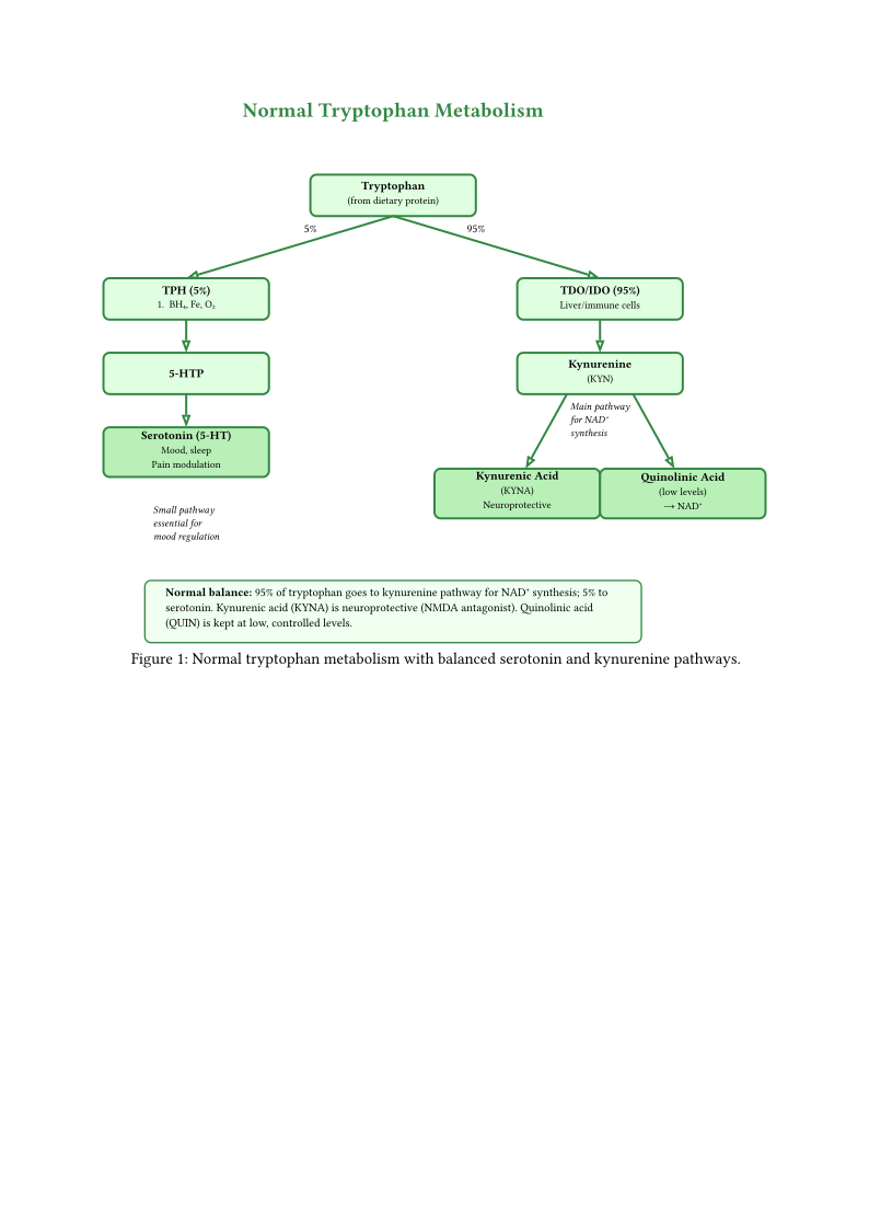
Kynurenine Pathway Dysregulation The kynurenine pathway metabolizes approximately 95% of dietary tryptophan and produces metabolites with diverse neuroactive effects. Quinolinic acid, an NMDA receptor agonist and excitotoxin, may contribute to neuroinflammation and cognitive dysfunction when elevated (Davis et al. 2023). Kynurenic acid, an NMDA receptor antagonist with neuroprotective properties, can become imbalanced with quinolinic acid, disrupting glutamatergic neurotransmission. Additionally, 3-hydroxykynurenine generates reactive oxygen species, potentially contributing to oxidative stress.
Immune activation, particularly interferon-gamma, stimulates the kynurenine pathway, providing a link between the immune abnormalities and neurological symptoms observed in ME/CFS (Mohsen Dehhaghi et al. 2022) (Kavyani et al. 2022).
The glutamatergic dimension of kynurenine pathway dysregulation acquires additional significance in light of the DecodeME GWAS findings. The genome-wide significant loci include SHISA6 (excitatory synaptic transmission at glutamatergic synapses) and UNC13C (glutamatergic synaptic transmission), while CA10 and DCC are associated with neuronal development in brain regions governing autonomic regulation (Section Replication Status: Not Yet Replicated (By Design) of Chapter Genetic and Epigenetic Factors). This convergence of genetic evidence (constitutional vulnerability in glutamatergic circuits) with metabolomic evidence (kynurenine-mediated disruption of glutamate/GABA balance) suggests a two-hit model: genetically vulnerable glutamatergic circuits are tipped into dysfunction by infection-driven quinolinic acid accumulation, and the resulting excitatory/inhibitory imbalance is maintained by chronic low-grade neuroinflammation. The Glutamatergic–Autonomic Bridge hypothesis (Chapter Integrative Models and Multi-System Pathophysiology, Speculation Integrative Speculations) formalises this model and identifies glutamatergic modulators (memantine, riluzole) as testable therapeutic candidates.
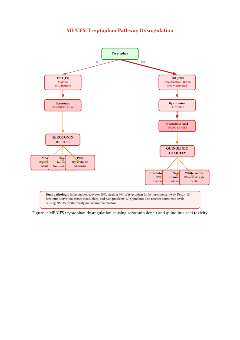
Figures tryptophan normal and tryptophan mecfs illustrate tryptophan metabolism dysregulation in ME/CFS. While normally approximately 95% of tryptophan is metabolized via the kynurenine pathway, inflammation-driven IDO overactivation can substantially increase this proportion. If kynurenine pathway flux increases to approximately 99% (a plausible estimate based on the magnitude of IDO upregulation observed in inflammatory conditions), this seemingly modest 4 percentage-point shift would dramatically reduce serotonin-available tryptophan from 5% to 1%—an 80% reduction in serotonin precursor availability—while quinolinic acid accumulation reaches toxic levels.
Serotonin Synthesis Diversion of tryptophan into the kynurenine pathway reduces availability for serotonin synthesis. Under conditions of IDO-driven inflammatory activation, up to 90% of tryptophan is catabolized through the kynurenine pathway (Mohsen Dehhaghi et al. 2022) (Mona Dehhaghi et al. 2022), leaving substantially less for serotonin synthesis. The estimated 80% reduction in serotonin-available tryptophan (derived from IDO upregulation magnitude documented in inflammatory states) may contribute to sleep disturbances, mood symptoms, pain amplification, and cognitive impairment observed in ME/CFS.
Enterochromaffin-Vagal Pathway: Gut-Serotonin-Brain Connection
Emerging research by Wirth and Scheibenbogen (2025) (Wirth and Scheibenbogen 2025) provides a mechanistic framework linking gut dysbiosis to central serotonergic dysfunction through the enterochromaffin-vagal pathway. This pathway offers a novel perspective on serotonin regulation in ME/CFS:
Certainty: 0.40. The enterochromaffin-vagal pathway may represent a critical gut-brain communication axis disrupted in ME/CFS. Enterochromaffin cells in the gut epithelium synthesize and release serotonin in response to mechanical stimulation and short-chain fatty acids (particularly butyrate). This serotonin activates 5-HT3 receptors on vagal afferent neurons, providing tonic input to the brainstem. In ME/CFS, gut dysbiosis and reduced butyrate production may impair enterochromaffin serotonin synthesis, leading to reduced vagal afferent signaling and downstream autonomic dysfunction.
Supporting evidence:
- Gut dysbiosis with reduced butyrate-producing bacteria is well-documented in ME/CFS (Giloteaux et al. 2016) (Hsu et al. 2025)
- Enterochromaffin cells require butyrate as a substrate for serotonin synthesis (Barton et al. 2025)
- Vagal tone (measured by HRV) is consistently reduced in ME/CFS patients (Escorihuela et al. 2020) (Walitt, Singh, LaMunion, Hallett, et al. 2024)
- The pathway explains how gut dysfunction could contribute to autonomic symptoms
Implications:
- Provides mechanistic link between gut dysbiosis and autonomic dysfunction
- Suggests butyrate supplementation and vagal stimulation as complementary therapies
- Explains why some ME/CFS patients have gastrointestinal symptoms alongside autonomic dysfunction
- Offers testable predictions for future research (e.g., butyrate supplementation improving HRV)
Limitations:
- Preprint status (not yet peer-reviewed)
- Enterochromaffin serotonin release not directly measured in ME/CFS
- Causality not established (could be reverse: vagal dysfunction → dysbiosis)
- Peripheral vs. central serotonin are distinct pools with different functions
4.3 Serotonergic Dysfunction
Beyond tryptophan diversion, multiple lines of evidence indicate primary serotonergic abnormalities in ME/CFS. PET imaging has demonstrated reduced serotonin transporter (5-HTT) density in the rostral anterior cingulate cortex (Yamamoto et al. 2004). Additional findings include abnormal responses to serotonergic challenge tests, correlations between serotonin markers and fatigue severity, and variable responses to serotonergic medications. The serotonergic system’s role in regulating sleep, mood, pain perception, and autonomic function positions it as a plausible contributor to the multisystem dysfunction of ME/CFS.
A preclinical model by Lee et al. (2024) provides complementary evidence from the opposite direction: rather than serotonin depletion via kynurenine diversion, serotonergic hyperactivity may also produce ME/CFS-like pathophysiology (J.-S. Lee et al. 2024). Using high-dose fluoxetine to induce elevated serotonin in mouse dorsal raphe nuclei and hypothalamus, the study demonstrated that chronic serotonergic excess produced severe fatigue, enhanced pain sensitivity, anxiety-like behaviour, and HPA axis dysfunction. The mechanism involved functional loss of 5-HT1A autoreceptors—the inhibitory feedback receptors that normally limit serotonin release—rather than quantitative receptor changes. This autoreceptor desensitisation amplified serotonergic signalling in limbic regions, producing a hyperserotonergia that paradoxically mimics the symptom profile attributed to serotonin deficiency in other models.
Certainty: 0.25. The coexistence of evidence for both serotonin depletion (tryptophan diversion via kynurenine pathway) and serotonin excess (5-HT1A desensitisation) in ME/CFS models suggests that serotonergic dysfunction may be bidirectional rather than unidirectional. Different compartments or brain regions may experience opposite perturbations simultaneously: peripheral serotonin pools depleted by IDO-driven tryptophan diversion while central raphe-limbic circuits become hyperactive due to 5-HT1A autoreceptor dysfunction. If correct, this explains the heterogeneous and sometimes paradoxical responses of ME/CFS patients to serotonergic medications (SSRIs helping some patients while worsening others). This remains a preclinical observation in mice; direct measurement of regional serotonergic tone in ME/CFS patients has not been performed.
4.4 Dopaminergic Dysfunction
Dopamine abnormalities extend beyond the CSF catecholamine findings to include measurably reduced basal ganglia activation during reward-processing tasks (Miller et al. 2014). Functional MRI shows that reduced activation of the right caudate nucleus and globus pallidus correlates significantly with mental fatigue severity (\(r^2 = 0.49\), \(p = 0.001\)). The overlap between ME/CFS fatigue and the motivational symptoms observed in dopaminergic disorders (Parkinson’s disease, interferon-induced fatigue) supports a shared mechanism of inflammatory cytokine-mediated disruption of basal ganglia dopamine availability. A further, independent human signal comes from neuroendocrine challenge testing: ME/CFS patients mount an exaggerated prolactin response to buspirone — a probe that blocks the dopamine-D2 brake on the lactotroph — despite normal baseline prolactin (Sharpe et al. 1996), and a related d-fenfluramine challenge found prolactin responses lowest in depression and highest in CFS (Cleare et al. 1995), though the buspirone-versus-depression comparison itself has never been performed (see Chapter Endocrine and Metabolic Dysfunction The Prolactin Axis and the Buspirone Challenge). This D2-level finding is consistent with reduced activity in the tuberoinfundibular dopaminergic system as one candidate contributor to the broader dopaminergic picture documented in this section — a compartment (hypothalamic/pituitary) that the CSF metabolite indices above do not measure, and distinct from the striatal terminals probed by VMAT2-PET; whether these dopaminergic subsystems co-vary in ME/CFS is untested.
The striatum’s role in central fatigue was theorised over 25 years ago by Chaudhuri and Behan, who proposed that central fatigue results from failure in the integration of limbic input and motor functions within the basal ganglia, specifically through dysfunction of the striatal-thalamic-frontal cortical system (Chaudhuri and Behan 2000). This “symptom signalling” hypothesis — that heterogeneous upstream pathology (immune, metabolic, infectious) converges on striatal circuits to generate the experience of fatigue — has gained substantial genetic support from the cell-type enrichment findings described above (Achievement Cell-Type Enrichment Analyses Converge on Neuronal Signal). The DecodeME data now provide the first genetic evidence that the striatum’s medium spiny neurons are specifically implicated in ME/CFS risk, converting a 25-year-old neurological hypothesis into a testable genetic model.
Certainty: 0.72 (Long COVID structural); indirect support in ME/CFS — functional imaging 0.55, treatment response 0.45 (distinct evidence types, not pooled). Liu et al. (2026) used (+)[“11C”]DTBZ PET — an index of vesicular monoamine transporter 2 (VMAT2) density and a validated marker of dopaminergic nerve terminal integrity — in 24 long COVID patients vs 24 age-matched controls (Liu et al. 2026). VMAT2 binding was significantly reduced across all striatal subregions: ventral striatum (20% reduction), dorsal putamen (16%), and dorsal caudate (17%) (LME group effect P=4×10⁻⁵). The absolute percentage reduction is similar to early Parkinson’s disease VMAT2 reductions, though the functional threshold for classical PD motor symptoms is substantially higher (50–60% striatal DA loss); the clinical significance of a 16–20% VMAT2 reduction in a non-parkinsonian population is uncertain. The same Toronto cohort separately demonstrated elevated TSPO PET (microglial activation) (Braga et al. 2023) and MAO-B PET (astrogliosis) (Braga et al. 2025) in the striatum, establishing that gliosis and VMAT2 loss co-occur in the same patients — though whether this association is causal, epiphenomenal, or coincidental is unresolved. A COVID-recovered control group (n=24) showed normal VMAT2 binding indistinguishable from never-COVID controls (P=0.77), confirming that reduced VMAT2 is specific to persistent illness rather than a generic effect of SARS-CoV-2 exposure.
VMAT2 loss correlated with clinically meaningful symptoms: ventral striatum loss with apathy (r=−0.54, P=0.0069), dorsal putamen loss with motor slowing on finger tapping (r=0.51, P=0.010), and dorsal caudate loss with delayed verbal memory (r=0.58, P=0.0029). There was no correlation between VMAT2 binding and depression scores (BDI), suggesting symptom correlations are not driven by mood. Peripheral blood markers of dopamine metabolism (DOPAC, HVA) and neurofilament light chain (NfL) showed no relationship to striatal VMAT2 binding, consistent with the known dissociation between peripheral and central dopaminergic compartments.
Independent functional imaging provides the first evidence of disrupted resting-state connectivity in dopamine-associated striatal circuits in an actual ME/CFS cohort: Inderyas et al. (2026) used ultra-high-field 7T fMRI to show altered functional connectivity in the nucleus accumbens — a striatal hub strongly implicated in reward and motivation — and related circuits in ME/CFS (n=32) and long COVID (n=19) patients, with striatal-cerebellar-amygdala connectivity correlating with cognitive symptom scores (Inderyas et al. 2026). This complements the structural VMAT2-terminal finding (Liu) from a different direction: functional disruption in dopamine-associated circuits within an ME/CFS cohort argues that dopaminergic-system involvement is not confined to the SARS-CoV-2-triggered subset. The parallel is strengthened further by a randomized trial of a dual norepinephrine–dopamine reuptake inhibitor (solriamfetol) in ME/CFS, which improved fatigue severity (p=0.039) and the global executive composite (p=0.012) (Young et al. 2025) — providing preliminary evidence that catecholamine-signalling augmentation may be clinically useful, though post-exertional malaise was not assessed and the drug’s dual mechanism does not isolate a dopamine-specific effect. Caution applies: connectivity (BOLD) fMRI does not measure dopamine itself and Inderyas indexed functional coupling, not terminal density; a direct VMAT2-PET measurement in non-COVID ME/CFS has not yet been performed — the structural-to-functional bridge in ME/CFS therefore remains inferred rather than directly observed.
Consequence: If replicated in ME/CFS, this would establish dopaminergic terminal loss as a measurable, regionally specific neuropathology — providing a treatment target (dopamine augmentation) and a PET-based biomarker for patient stratification in clinical trials. Direct structural VMAT2-PET data in ME/CFS are still absent; the finding is structurally confined to long COVID and requires direct replication in ME/CFS cohorts. The indirect functional evidence in an ME/CFS cohort (Inderyas 2026 (Inderyas et al. 2026)) and the positive ME/CFS solriamfetol RCT ((Young et al. 2025)) justify pursuing that replication but do not substitute for it.
Falsifiable predictions. If VMAT2 loss is causal for apathy and motor slowing: (1) dopamine augmentation (L-DOPA) should improve apathy and motor speed in VMAT2-characterised long COVID patients; (2) longitudinal VMAT2 PET should show signal stability over 12 months (consistent with structural loss, not activity-dependent fluctuation). Falsified if dopamine augmentation fails to improve symptoms in VMAT2-low patients in a randomised trial, or if the VMAT2-apathy correlation is not replicated in an independent cohort.
Limitations. Long COVID only (no ME/CFS data). Study sample was young (mean 32.2 years), apathy-predominant (all participants had apathy by inclusion), and n=24 limits subgroup analyses. VMAT2 binding is a proxy for terminal integrity but could theoretically reflect reduced vesicle density per terminal rather than terminal loss per se — though treatment implications may be similar in either case. The absence of pre-COVID baseline prevents definitive attribution of VMAT2 reduction to COVID-19 rather than a pre-existing trait; however, COVID-recovered controls with normal VMAT2 argue against this interpretation. An alternative interpretation is that [11C]DTBZ binding is sensitive to vesicular pH gradient, which could be reduced by low striatal ATP (mitochondrial dysfunction) without any change in VMAT2 protein or terminal number — a confound not excluded by the study design. Moreover, the findings may be SARS-CoV-2-specific (given the virus’s DA neuron tropism via ACE2 (Chen et al. 2020) (Yang et al. 2024); an animal model further shows SARS-CoV-2 can invade the substantia nigra pars compacta where dopaminergic cell bodies reside (Pokharel et al. 2025)) and not generalize to ME/CFS triggered by EBV, HHV-6, or other pathogens. Aregawi et al. (2026) found normal CSF dopamine pathway indices (DA + DOPAC + HVA) in PI-ME/CFS with only noradrenergic deficiency (Aregawi et al. 2026) — a finding that is inconsistent with functionally significant global DA terminal loss. This observation currently lacks a resolution: it may indicate that VMAT2 loss does not generalise to PI-ME/CFS, that terminal loss is compensated by upregulated per-terminal synthesis, or that CSF DA metabolites do not reflect striatal VMAT2 status. Simultaneous CSF+PET measurement in the same patients is required to adjudicate.
Certainty: n/a. The VMAT2-apathy association (r=−0.54) in Liu et al. (2026) is cross-sectional. An alternative causal model is equally consistent with the data: neuroinflammation (TSPO + MAO-B PET elevations in the same cohort) produces sickness behaviour and apathy via cytokine signalling, which reduces behavioural activation, which in turn downregulates VMAT2 through use-dependent plasticity — making VMAT2 reduction a consequence rather than a cause of symptoms. Under this model, dopamine augmentation would target a biomarker rather than the driving mechanism, and anti-inflammatory or behavioural activation approaches would be more appropriate. The evidence cannot currently distinguish these interpretations: correlation does not establish causal direction.
Consequence: If VMAT2 loss is epiphenomenal, the entire dopamine augmentation treatment framework targets the wrong mechanism — anti-inflammatory strategies would be the correct intervention. The single decisive test is a dopamine augmentation trial in VMAT2-characterised patients: if DA agents improve symptoms in VMAT2-low but not VMAT2-normal patients, causality is supported. If no differential response, the epiphenomenon interpretation is strengthened.
Testable predictions. A randomised controlled trial of L-DOPA vs placebo in VMAT2-low long COVID patients: (a) if VMAT2 is causal, apathy and motor slowing improve with L-DOPA and baseline VMAT2 binding predicts response magnitude; (b) if VMAT2 is epiphenomenal, L-DOPA produces no differential benefit in VMAT2-low vs VMAT2-normal groups, or worsens symptoms via orthostatic/oxidative side effects.
Certainty: n/a. The Liu et al. (2026) VMAT2 PET finding is in long COVID — a condition with established SARS-CoV-2 tropism for dopaminergic neurons via ACE2 receptors expressed in substantia nigra and ventral tegmental area (Chen et al. 2020) (Liu et al. 2026). Whether other ME/CFS triggers (EBV, HHV-6, Borrelia, physical trauma, surgery) produce comparable dopaminergic terminal loss is unknown. The single existing CSF study in PI-ME/CFS found normal dopamine pathway metabolites (Aregawi et al. 2026), arguing against a generalised DA deficiency. However, ultra-high-field 7T fMRI now shows reduced nucleus accumbens functional connectivity in a mixed ME/CFS cohort (Inderyas et al. 2026 (Inderyas et al. 2026)), whose trigger aetiologies were not restricted to SARS-CoV-2 — providing preliminary functional (though not structural terminal-density) evidence that dopaminergic-circuit involvement may extend beyond COVID. Alphavirus models (Bantle 2019, Ross River/Sindbis/CHIKV) show similar DA neuron tropism (Bantle et al. 2019), and postencephalitic parkinsonism after 1918 influenza is well-documented, suggesting postviral DA vulnerability may extend beyond SARS-CoV-2 — but direct VMAT2 PET in non-COVID ME/CFS is still lacking.
Consequence: If VMAT2 loss is COVID-specific, the treatment implication (dopamine augmentation) applies only to the post-COVID ME/CFS subset and the field should pursue NE-targeted therapies for the broader ME/CFS population, consistent with Aregawi et al.’s selective noradrenergic findings. If VMAT2 loss generalizes across triggers, dopamine augmentation becomes a broadly applicable strategy.
Testable predictions. VMAT2 PET in non-COVID PI-ME/CFS (n≥20 EBV-onset, n≥20 other triggers) will show either (a) reduced binding equivalent to Liu 2026 → mechanism is general postviral; (b) normal binding → mechanism is SARS-CoV-2-specific.
Section “Striatal Neuron Diversity: Eccentric Medium Spiny Neurons” (below) examines cellular diversity within the striatum, including eccentric medium spiny neurons (eMSN) that integrate D1/D2 dopamine signaling—a cell type potentially relevant to this dopaminergic dysfunction.
Certainty: 0.15. VMAT2 is the sole transporter packaging dopamine into synaptic vesicles, protecting against cytosolic dopamine oxidation. Reduced VMAT2 → more cytosolic dopamine → spontaneous auto-oxidation to dopamine-quinones → Complex I inhibition and reactive oxygen species generation. In a patient with pre-existing mitochondrial vulnerability (documented in ME/CFS — reduced ATP/ADP ratio, 272-gene metabolic module dysfunction), this creates a feed-forward toxicity loop: VMAT2 loss → oxidative dopamine damage → further Complex I impairment → more ATP depletion → further VMAT2 dysfunction (ATP is required for the vesicular proton gradient). This synergy explains why even a 16–20% VMAT2 reduction could produce disproportionate functional impact — the oxidative burden transforms a modest storage deficit into an escalating energy crisis (Liu et al. 2026).
Consequence: If correct, dopamine augmentation (L-DOPA) could transiently worsen symptoms by flooding a compromised system with oxidation-prone cytosolic dopamine — implying that treatment must be paired with antioxidant support and mitochondrial protection. (Origin: brainstorm.)
Falsifiable predictions. CSF 5-S-cysteinyl-dopamine (marker of dopamine oxidation) should be elevated in patients with VMAT2 loss and correlate inversely with Complex I activity in platelets. L-DOPA challenge should show a biphasic response: initial worsening of fatigue and cognitive symptoms (from oxidation) followed by improvement as surviving terminals clear the cytosolic load.
Limitations. Dopamine-quinone formation has not been measured in long COVID or ME/CFS. The ΔpH sensitivity of [11C]DTBZ binding means the PET signal could reflect ATP-dependent vesicular pH rather than terminal density — which would actually strengthen this model (VMAT2 binding as a readout of the energy crisis rather than structural loss).
Certainty: 0.25. VMAT2 density can modulate quantal size — the amount of dopamine per released vesicle. A 16–20% VMAT2 reduction predicts approximately 20% less dopamine released per action potential. Effort-based decision-making depends on phasic dopamine bursts during activity; each activity produces less dopamine-mediated reinforcement. Over time, the brain’s cost-benefit computation shifts: effort is perceived as increasingly costly because the “this was worthwhile” signal is persistently attenuated (Liu et al. 2026). Patients become trapped in a downward spiral — less activity → less dopamine release → weaker reinforcement → even less motivation — despite the desire to be active.
This model is consistent with the basal ganglia effort-cost miscalibration hypothesis (Speculation Basal Ganglia “Effort Cost” Miscalibration Hypothesis) and provides a neurochemical substrate for the behavioral observations: the striatum does not miscompute effort cost — it receives a genuinely weaker reward signal per unit of effort, making the correct computation produce an apparently inflated cost.
Consequence: This reframes the apathy of postviral illness as a biochemically driven phenomenon — reduced dopamine per action potential means each activity produces weaker reinforcement, creating a rational motivational deficit from a biological constraint. (Origin: brainstorm.)
Falsifiable predictions. [11C]raclopride displacement PET during reward tasks should show blunted dopamine release in long COVID patients with VMAT2 loss. Effort discounting tasks should show elevated discounting rates (choose low-effort/low-reward over high-effort/high-reward) correlating with VMAT2 signal. Dopamine augmentation (L-DOPA) should normalize effort discounting behavior.
Limitations. No PET raclopride displacement data in long COVID or ME/CFS. Behavioral effort-discounting tasks have not been administered in VMAT2-characterized patients. The model assumes VMAT2 is the rate-limiting factor for phasic DA release, but firing rate, synthesis capacity, and D2 autoreceptor regulation also modulate DA output.
Certainty: 0.35. Capuron et al. (2012) demonstrated that interferon-alpha (IFN-α) therapy — used in hepatitis C and cancer treatment — produces a syndrome of apathy, psychomotor slowing, and fatigue that closely mirrors the neuropsychiatric profile of long COVID and ME/CFS (Capuron et al. 2012). PET imaging shows IFN-α reduces ventral striatal dopamine release via p38 MAPK and IDO/kynurenine pathway activation, which suppresses tyrosine hydroxylase expression and reduces dopamine synthesis. This is the best-characterized human model of peripherally triggered central dopamine dysfunction.
The model converges with the VMAT2 PET finding in a key respect: the symptom profile (apathy > mood disturbance, motor slowing > subjective fatigue) is identical whether the upstream cause is IFN-α signal transduction (cytokine-driven DA synthesis failure) or SARS-CoV-2 infection of DA neurons (DA terminal loss). The convergence on a shared downstream dopamine phenotype suggests that DA augmentation should benefit both groups, and that existing trial designs from IFN-α studies can be adapted for long COVID and ME/CFS (Capuron et al. 2012). Aregawi et al. (2026) normal CSF DA pathway finding in PI-ME/CFS does not conflict with this model, since IFN-α-induced dopamine dysfunction also occurs via regulatory mechanisms that may not reduce total CSF metabolite output (Aregawi et al. 2026).
Consequence: A rich clinical research literature — including neuroimaging biomarkers (DAT SPECT, raclopride displacement PET) and interventional trial designs validated in IFN-α populations — can be directly adapted for biomarker-stratified dopamine augmentation trials in long COVID and post-infectious ME/CFS. This substantially lowers the translational barrier. (Origin: brainstorm.)
Limitations. IFN-α is an iatrogenic model, not a natural postviral disease. The mechanism is synthesis suppression (TH downregulation) rather than terminal loss (VMAT2 reduction) — the downstream phenotype converges, but the upstream biology differs. No DA augmentation trial in IFN-α-treated patients has been conducted in ME/CFS or long COVID populations.
Research directions. The strongest priority experiments are: (1) VMAT2 PET with paired CSF DA metabolite sampling to reconcile Liu 2026 and Aregawi 2026 in the same patients; (2) DaTSCAN SPECT ([123I]FP-CIT) in long COVID as a widely accessible alternative to VMAT2 PET — if DAT binding parallels VMAT2 loss, clinical translation is immediate; (3) a DA augmentation trial in VMAT2-characterised long COVID patients, adapting IFN-α trial methodology, with pre- and post-treatment apathy and motor speed outcomes.
Falsifiable predictions. If the IFN-α and long COVID VMAT2 models converge on shared dopaminergic deficiency: (1) [18F]FDOPA PET should distinguish the two mechanisms — preserved dopamine synthesis (normal FDOPA Ki) with reduced VMAT2 binding in long COVID versus reduced FDOPA Ki in IFN-α-treated patients; (2) VMAT2 PET in IFN-α-treated patients who develop apathy should show normal binding (since the mechanism is synthesis suppression, not terminal loss). Falsified if VMAT2 PET is abnormal in IFN-α patients, or if [18F]FDOPA PET is abnormal in long COVID patients with VMAT2 loss.
Certainty: 0.50. Several evidence-quality concerns constrain the VMAT2-long COVID findings and their extrapolation to ME/CFS: (a) The Liu et al. (2026) study is n=24 per group — adequate for PET but susceptible to inflated effect sizes from small-n sampling; independent replication in a larger, multicentre cohort is essential before strong conclusions. (b) The Toronto clinic is a tertiary referral centre, potentially enriching for severe, neurologically-predominant long COVID phenotypes; the 16–20% VMAT2 reduction may not generalise to community-based long COVID populations. (c) The correlation between VMAT2 loss and apathy (r=−0.54) could be reverse-causal: apathy → reduced activity → use-dependent VMAT2 downregulation, rather than VMAT2 loss causing apathy. Longitudinal PET after behavioural activation could distinguish these directions. (d) The reported p-values were not corrected for the full multiple-comparison burden (multiple subregions, multiple clinical tests, multiple cognitive measures); the strongest associations (memory r=0.58) would likely survive correction, but weaker subregion-specific claims should be interpreted cautiously. (e) The 16–20% VMAT2 loss is well below the 50–60% threshold at which Parkinson’s disease motor symptoms emerge — while apathy and cognitive slowing can occur at lower DA loss levels (20–30% in aging and prodromal PD), it remains possible that the VMAT2 signal is a clinically silent epiphenomenon downstream of neuroinflammation, with symptoms driven by non-DA mechanisms. Only a dopamine augmentation trial in VMAT2-characterised patients can resolve whether the loss is symptomatically relevant. (f) A purely methodological concern: VMAT2 normally declines 5–8% per decade after age 40; a 2–3 year age difference between groups could partially confound the signal, though the COVID-recovered controls with normal VMAT2 and the age-matching (±5 years) mitigate this concern. (g) The possibility that VMAT2 loss reflects broad TRPV1+ neuronal damage rather than DA-specific pathology cannot be excluded from the current data.
Consequence: The VMAT2-long COVID finding is a strong candidate mechanism for apathy and motor slowing in a specific phenotype of long COVID, but its extrapolation to ME/CFS requires cautious framing until replication, VMAT2 PET in ME/CFS, and a causal test (DA augmentation trial) are available. The evidence base supports integration as observation and speculation with explicit uncertainty — not as established ME/CFS pathophysiology. (Origin: brainstorm — Phase 5 evidence quality consolidation.)
Chapter reference. Observation Striatal Dopaminergic Terminal Loss Quantified by VMAT2 PET in Long COVID, Speculations DA-ATP “Double-Hit” — VMAT2 Loss Synergises with Mitochondrial Dysfunction and Activity-Dependent DA Release Failure — A Biological “Motivation Trap”.
Evidence base. Liu et al. (2026) VMAT2 PET (Observation Striatal Dopaminergic Terminal Loss Quantified by VMAT2 PET in Long COVID) demonstrates 16–20% striatal VMAT2 binding reduction in long COVID, with co-occurring TSPO PET (microglial activation) and MAO-B PET (astrogliosis) from the same Toronto patient sample — three multi-modal signals from one cohort that show association, not independently replicated convergence (Liu et al. 2026) (Braga et al. 2023) (Braga et al. 2025). Postmortem evidence confirms SARS-CoV-2’s capacity to directly infect DA neurons via ACE2 and induce senescence (Yang et al. 2024). The clinical correlations (apathy r=−0.54, motor slowing r=0.51, memory r=0.58) map onto functionally distinct striatal subregions, and the IFN-α human model (Observation Interferon-α-Induced Depression as a Human Model of Cytokine-Driven Dopamine Suppression) provides convergent evidence that cytokine-mediated dopamine dysfunction produces an identical symptom profile via a different upstream mechanism (synthesis suppression rather than terminal loss) (Capuron et al. 2012).
The mechanism has three interacting components: (1) Structural: SARS-CoV-2 infection or gliosis-driven damage reduces VMAT2-positive dopaminergic terminals, lowering vesicular dopamine storage capacity; (2) Energetic: reduced VMAT2 increases risk of cytosolic dopamine oxidation (Speculation DA-ATP “Double-Hit” — VMAT2 Loss Synergises with Mitochondrial Dysfunction), creating a feed-forward loop with mitochondrial dysfunction — each compound exacerbates the other; (3) Functional: reduced quantal dopamine release per action potential attenuates the brain’s “effort was worthwhile” signal (Speculation Activity-Dependent DA Release Failure — A Biological “Motivation Trap”), shifting the cost-benefit computation of every activity toward the “not worth it” outcome.
The treatment implication (Speculation Dopaminergic Augmentation for Apathy-Predominant and Motor-Slowing Phenotypes in Post-Infectious ME/CFS, ch28, with comprehensive safety assessment at Warning Safety Assessment for Dopaminergic Agents in ME/CFS) is that dopamine augmentation — L-DOPA to increase synthesis, MAO-B inhibitors to reduce degradation, or dopamine agonists to bypass terminals — targets the functional deficit directly. However, the structural component (terminal loss) may be irreversible, limiting ceiling effects, and the energetic component warns that augmenting cytosolic dopamine without adequate antioxidant or mitochondrial support could worsen oxidative damage.
Key constraint: This model is structurally derived from long COVID evidence. Direct VMAT2-PET terminal-density data in ME/CFS do not yet exist. Functional imaging of dopaminergic circuits in an actual ME/CFS cohort (reduced nucleus accumbens functional connectivity, Inderyas et al. 2026 (Inderyas et al. 2026)) and a positive solriamfetol RCT in ME/CFS ((Young et al. 2025)) now provide indirect support that dopaminergic-system involvement extends beyond the SARS-CoV-2-triggered subset. Aregawi et al. (2026) normal CSF DA pathway indices in PI-ME/CFS (Aregawi et al. 2026) still constrain the generality — preserved synthesis capacity could compensate for reduced terminal density. The model may apply most strongly to the SARS-CoV-2-triggered subset of post-infectious ME/CFS (Open Question Does VMAT2 Loss Generalize Beyond SARS-CoV-2 to Other ME/CFS Triggers?), with the functional and therapeutic ME/CFS evidence warranting direct structural replication.
Consequence: If validated in ME/CFS, this model would shift treatment from empiricism to mechanism-matching for a specific patient phenotype (apathy-predominant post-infectious illness with objective motor slowing), using VMAT2 PET or DAT SPECT as a stratification biomarker. The most decisive single experiment is VMAT2 PET in non-COVID PI-ME/CFS — negative result confines the model to post-COVID ME/CFS; positive result establishes a general postviral dopaminergic vulnerability.
4.5 Striatal Neuron Diversity: Eccentric Medium Spiny Neurons
Medium spiny neurons (MSNs) are the dominant neuronal population in the striatum (caudate nucleus, putamen, and nucleus accumbens) and are critical for basal ganglia function in motor control, reward processing, and effort allocation decisions. MSNs are classically divided into D1 receptor-expressing (direct pathway) and D2 receptor-expressing (indirect pathway) subtypes, but recent discoveries have revealed unexpected cellular diversity within this system.
Certainty: 0.85. He et al. (2021) identified nine distinct MSN subtypes in rhesus macaque striatum through single-nucleus RNA-seq, including a previously overlooked D1/D2 hybrid population termed “eccentric” medium spiny neurons (eMSN) (He et al. 2021). These hybrid neurons express both DRD1 and DRD2 dopamine receptors, are marked by high RXFP1 expression, constitute approximately 10% of MSNs in the dorsal striatum, and are uniformly distributed throughout this region. The eMSN represent a distinct supercluster with a unique genetic profile differentiating them from classical D1- and D2-MSNs. This discovery was validated by fluorescence in situ hybridization (FISH) confirming co-expression of DRD1, DRD2, and RXFP1 in individual neurons. Study: (n=4 rhesus macaques, snRNA-seq + FISH validation, Current Biology, certainty: High, replicated in mice, human data emerging).
Certainty: 0.95. Siletti et al. (2023) generated a comprehensive single-nucleus RNA-seq atlas of the entire human brain (3.3 million cells from 105 dissections across 3 donors) and confirmed eMSN as a distinct human cell type (Siletti et al. 2023). The Human Protein Atlas lists “eccentric medium spiny neurons” as a cluster with marker genes GAD1, GAD2, CXCL14, DRD1, and ADARB2, establishing eMSN as a validated component of human striatal circuitry. This landmark Science publication provides the definitive reference for human brain cell-type diversity. Study: (n=3 donors, 3.3M cells, snRNA-seq, Science, certainty: Very High, dataset publicly available for validation).
Functional implications of striatal neuron diversity.
The existence of eMSN with hybrid D1/D2 receptor expression challenges the classical direct/indirect pathway dichotomy. By integrating both dopamine receptor types, eMSN may serve a unique integrative function in basal ganglia circuitry—potentially modulating conflict resolution, effort-based decision-making, or motivational state evaluation. Their uniform distribution in the dorsal striatum suggests a broad role in motor and cognitive functions rather than a region-specific specialization.
The dopamine receptor hybridity and RXFP1 expression (RXFP1 encodes relaxin-family peptide receptor 1, linked to neuroprotection and neuroinflammation modulation) raise the possibility that eMSN have distinct physiological properties affecting stress responses, neuroinflammation, or metabolic regulation. However, the specific functional role of eMSN in human brain physiology remains largely unknown, and no studies have examined eMSN in disease states including ME/CFS.
Certainty: 0.70. Multiple independent post-GWAS enrichment analyses applied to DecodeME and Million Veteran Program data converge on a single finding: ME/CFS genetic risk maps to neuronal cell types in the CNS, not to immune cells. Cell-type enrichment analysis using the Siletti et al. 2023 Human Brain Atlas (461 cell types) via the Duncan et al. 2025 pipeline identified medium spiny neurons (MSNs), particularly the eccentric MSN (eMSN) subtype, as the most specific cell-type hit for ME/CFS (Maccallini 2026) (Duncan et al. 2025) (Siletti et al. 2023). Dropviz mouse brain atlas analysis independently identified striatal neurons (7 of 13 significant cell types) (Saunders et al. 2018). A third atlas (DESCARTES human fetal) found inhibitory interneuron enrichment (J. H. Lee et al. 2026). Meanwhile, Finucane’s stratified LDSC pipeline applied to ImmGen immune cell data — a reference dataset covering major immune cell types (T cells, B cells, NK cells, macrophages, dendritic cells) — found zero significant immune cell-type associations for ME/CFS (Finucane et al. 2018). This means ME/CFS common-variant genetic risk does not enrich in any immune cell type — the immune signal is null (Finucane et al. 2018). Rare variant analysis (Snyder et al. 2025) independently implicates neuronal genes in synaptic function, complementing common variant findings (Snyder, Zhao, et al. 2025).
The broad neuronal signal (brain tissue enrichment, gene-set enrichment) is robust (certainty 0.80). Finer cell-type resolution — particularly the eMSN specificity — is method-dependent (Brouwer 2026 review: fine cell-type enrichment varies with pipeline parameters). MSN enrichment is also not unique to ME/CFS: Duncan et al. 2025 found MSNs enriched for schizophrenia, depression, sleep duration, and alcohol consumption. Nevertheless, the data firmly establish that ME/CFS has a heritable component converging on neuronal gene expression, providing genetic evidence for a CNS component.
Consequence: This genetic architecture — risk mapped to neurons, not immune cells — defines ME/CFS as a disease with a CNS genetic substrate. For researchers: prioritises neuronal mechanisms as direct readouts of genetic risk rather than downstream effects. For patients: confirms that ME/CFS has a brain-based genetic component, countering the narrative that it is primarily psychological or behavioural.
Study: (meta-analysis n=19,470 cases, 3 independent cell atlases, 2 analytic methods, convergent with rare variant analysis; key caveat: eMSN resolution is method-dependent).
Certainty: 0.40. Chaudhuri and Behan (2000) proposed that central fatigue results from failure of the “non-motor function” of the striatal-thalamic-frontal cortical system (Chaudhuri and Behan 2000). In this model, heterogeneous upstream pathology (immune activation, metabolic stress, infection-triggered damage) converges on striatal circuits that generate the experience of fatigue, malaise, and effort intolerance — the striatum acts as a common bottleneck where diverse biological insults produce a similar symptom output.
The cell-type enrichment findings described above (Achievement Cell-Type Enrichment Analyses Converge on Neuronal Signal) provide the first genetic evidence consistent with this framework: if dozens of upstream pathologies converge on MSN function, MSN-expressed genes would show genetic enrichment even though the primary pathology is elsewhere. This would explain (1) why brain enrichment is so strong despite no single brain pathology identified, (2) why the immune cell enrichment is null — immune pathology is upstream and acquired rather than genetically encoded in common variants, and (3) why MSN enrichment is shared across multiple brain-affecting traits (schizophrenia, depression, alcohol consumption, sleep duration — (Duncan et al. 2025)) — the striatum is a convergence point for numerous forms of brain dysfunction, not a disease-specific risk locus.
Falsifiable predictions. (1) Resting-state fMRI: striatal-thalamic-frontal connectivity explains significant additional variance in fatigue severity beyond peripheral inflammation markers (CRP, IL-6, TNF-alpha) in hierarchical regression. Falsified if striatal connectivity adds no significant incremental variance beyond peripheral markers (ΔR² ≈ 0), or if peripheral markers outpredict striatal connectivity in head-to-head comparison. (2) MSN-specific PRS vs whole-brain PRS: a PRS derived from SNPs enriched in MSN-expressed genes outperforms a whole-brain PRS in predicting fatigue severity within the ME/CFS population. Falsified if whole-brain PRS explains ≥ MSN-specific PRS variance in fatigue severity, or if neither PRS predicts fatigue severity (genetic architecture not striatal-specific).
Consequence: If correct, this model reframes ME/CFS as a disorder of a specific neural circuit rather than a vague “multisystem illness” — the diverse symptoms converge on striatal output, making the striatum a rational therapeutic target (dopamine modulation, neuromodulation). For patients: shifts the narrative from “it’s all in your head” to “your genetic risk is expressed in a specific brain circuit that we can study and potentially target.”
Limitations: Theoretical framework now with genetic support; no direct measurement of striatal-thalamic-frontal circuit function in ME/CFS; the convergence model is consistent with but not uniquely predicted by the genetic data — alternative explanations (e.g., MSNs as passive transcriptional tags, entirely separate aetiologies that happen to affect MSN gene expression) remain viable. Origin: literature synthesis (Chaudhuri & Behan 2000 + DecodeME cell-type enrichment).
Certainty: 0.85. While the broad neuronal enrichment signal across tissues and gene-sets is robust, finer cell-type resolution is method-dependent. The eMSN association varies with analysis pipeline parameters, LD reference panel choice, and gene window size (Brouwer 2026 comprehensive review of enrichment methodology). MSN/eMSN specificity is estimated at certainty 0.40–0.50, while the broader neuronal signal stands at certainty 0.80. Other cell types reach significance depending on which atlas is used: glutamatergic neurons in cerebellar white matter (Seeker 2023 dataset), inhibitory interneurons (DESCARTES fetal atlas). MSN enrichment is also shared across multiple brain-relevant traits (schizophrenia, depression, alcohol consumption, sleep duration — Duncan et al. 2025), so it cannot function as an ME/CFS-specific diagnostic marker.
These caveats do not undermine the core finding: across all methods and atlases, ME/CFS genetic risk maps to neurons, not immune cells. The unresolved question is which neuronal subtypes are causally involved, and whether the genetic signal points to MSNs specifically or to a broader neuronal process that MSNs happen to tag by their transcriptional profile.
Consequence: Patients and clinicians should understand that the genetic findings identify “brain cells, specifically neurons” as where risk is encoded, but the claim that a particular neuron type (medium spiny neurons in the striatum) is the key cell type should be treated as a working hypothesis, not an established result. The method-dependent nature of fine cell-type resolution means different analysis choices could highlight different neuronal populations — the field is converging on neuronal biology broadly, not on a single cell type.
Study: (3 atlases, 2 methods, convergent neuronal signal; fine resolution method-dependent).
No published studies have examined eMSN populations, gene expression, or functional activity in ME/CFS patients. The cell-type enrichment findings above (Achievement Cell-Type Enrichment Analyses Converge on Neuronal Signal) provide genetic evidence that eMSN-expressed genes are enriched for ME/CFS risk variants, but this does not establish whether eMSN are functionally dysregulated. Post-mortem ME/CFS brain tissue studies (including the Dutch autopsy findings described below) could examine eMSN markers (RXFP1, CXCL14, ADARB2) via immunohistochemistry or single-nucleus RNA-seq to determine whether eMSN density, distribution, or gene expression differs from healthy controls. Alternatively, functional MRI studies with eMSN-informed computational modeling could test whether eMSN-related circuit patterns predict symptom severity.
Testable predictions. Falsified if post-mortem brain tissue studies show normal eMSN density, distribution, and gene expression in ME/CFS patients compared to healthy controls. Falsified if fMRI studies with eMSN-informed computational modeling show no correlation between eMSN-related circuit patterns and ME/CFS symptom severity.
Consequence: none — basic mechanistic observation with no translational horizon. Functional characterisation of eMSN in ME/CFS would validate or refute the genetic enrichment findings at the protein/circuit level, but neither result directly changes patient care.
The cell-type enrichment analyses described above (Achievement Cell-Type Enrichment Analyses Converge on Neuronal Signal) have partially answered the previous open question: MAGMA cell-type analysis using the Human Brain Atlas does find eMSN/medium spiny neuron enrichment in ME/CFS (Maccallini 2026). However, convergent evidence from multiple atlases and analysis pipelines is needed to confirm whether the MSN/eMSN signal is genuinely causal or merely a transcriptional tag. Remaining narrower questions: (1) Does the eMSN enrichment survive independent replication with a fully independent GWAS (not MVP, which partially overlaps DecodeME cohorts)? (2) Do voxel-level brain eQTL colocalisation analyses (e.g., using PsychENCODE dorsolateral PFC eQTLs) confirm that SNP effect sizes at DecodeME loci correlate with eMSN marker gene expression specifically, rather than reflecting general neuronal transcription? (3) Does the MSN signal reflect a causal variant in MSNs themselves, or a variant in genes that are broadly expressed in neurons but that MSN transcriptomes happen to tag due to their high neuronal transcript diversity?
Testable predictions. Falsified if an independent GWAS replicates the brain tissue enrichment but does not identify eMSN/MSN as the top cell-type hit. Alternatively, supported if colocalisation analysis confirms eMSN-specific expression patterns at DecodeME genome-wide significant loci.
Certainty: 0.40. The striatum assigns effort cost to planned actions through basal ganglia circuitry that evaluates reward versus effort tradeoffs. In ME/CFS, basal ganglia circuits (potentially involving eMSN integration of D1/D2 signaling) may systematically overestimate physiological cost, leading to pre-emptive inhibition of motor activity and exaggerated subjective fatigue perception. This is not “laziness” or deconditioning but a miscalibrated cost-benefit computation at the neural circuit level.
Mechanistic rationale. Basal ganglia dysfunction correlates with mental fatigue severity (r²=0.49, p=0.001) (Miller et al. 2014). The striatum encodes effort-cost calculations via dopaminergic modulation: D1 receptor activation promotes “go” signals for high-effort/high-reward actions, while D2 receptor activation promotes “no-go” signaling for low-reward actions. eMSN with hybrid D1/D2 expression may normally provide nuanced integration of competing signals, but dysfunction could create pathological cost inflation—every action is perceived as excessively costly.
Testable predictions. ME/CFS patients will show abnormal striatal activation during effort-cost decision tasks (fMRI), overvaluing physiological costs relative to rewards. Computational modeling will reveal elevated cost-sensitivity parameters in patient decision-making. Dopaminergic agents that modulate striatal output will preferentially improve patients with prominent effort-initiation difficulty.
Falsified if: ME/CFS patients show normal striatal activation patterns during effort-cost decision tasks; computational modeling reveals cost-sensitivity parameters comparable to healthy controls; dopaminergic agents fail to preferentially improve patients with effort-initiation difficulty.
Treatment implications. Cognitive approaches targeting effort perception recalibration; dopaminergic agents that rebalance D1/D2 signaling; non-pharmacological effort-cost retraining protocols.
Limitations. No direct evidence linking eMSN to effort-cost computation; dopamine system dysregulation in ME/CFS is complex (both reduced central catecholamines and potential peripheral hyperactivity); effort-cost miscalibration may be epiphenomenon rather than cause.
Certainty: 0.35. The striatum integrates autonomic status with motor planning through basal ganglia-thalamus-cortex loops. In ME/CFS, eMSN dysfunction or broader striatal pathology may create a disconnect between autonomic state and motor command generation, leading to inappropriate activity initiation despite inadequate autonomic preparation (causing crashes) or excessive inhibition despite sufficient resources (contributing to disability).
Mechanistic rationale. Autonomic dysfunction is a primary driver of post-exertional malaise (PEM) in ME/CFS (Keller et al. 2024). Striatum modulates autonomic output via projections to brainstem nuclei controlling heart rate, blood pressure, and respiratory patterns. The temporal-parietal junction dysfunction documented in ME/CFS (Walitt, Singh, LaMunion, Hallett, et al. 2024) may impair integration of interoceptive signals (autonomic state) with striatal motor planning, creating a mismatch where motor commands proceed without autonomic preparation or resources are conserved despite adequate physiological capacity.
Testable predictions. ME/CFS patients will show abnormal striatal-heart rate variability coupling during postural changes. Striatal activity measured via fMRI will predict autonomic collapse events before they occur, providing a potential early warning biomarker. Patients with pronounced striatal-autonomic decoupling will experience more frequent and severe PEM episodes.
Falsified if: ME/CFS patients show normal striatal-heart rate variability coupling during postural changes; striatal fMRI activity does not predict autonomic collapse events; striatal-autonomic decoupling severity does not correlate with PEM episode frequency or severity.
Treatment implications. Autonomic biofeedback combined with striatal-targeted cognitive training; interventions that enhance interoceptive awareness and striatal-autonomic coupling; graded approaches that ensure autonomic readiness before motor activation.
Limitations. No direct evidence measuring striatal-autonomic coupling in ME/CFS; autonomic dysfunction has multiple potential drivers (peripheral, central, vascular); isolating striatal contribution to autonomic dysregulation experimentally is challenging.
4.6 Norepinephrine and the Locus Coeruleus
The locus coeruleus (LC), the primary source of brain norepinephrine, plays critical roles in arousal and sleep-wake regulation, attention and cognitive flexibility, stress responses, and autonomic nervous system modulation.
Certainty: 0.40. (0.35 to 0.40: Aregawi 2026 composite NE Pathway index provides additional supporting CSF evidence — partial replication, same NIH center but extended methodology.) LC dysfunction may explain the constellation of arousal, attention, and autonomic abnormalities in ME/CFS. The CSF catecholamine abnormalities documented in ME/CFS — including reduced DOPA, DOPAC, and DHPG (Walitt, Singh, LaMunion, Hallett, et al. 2024) and selectively reduced NE Pathway index (NE+DHPG+MHPG) with normal DA Pathway (DA+DOPAC+HVA) (Aregawi et al. 2026) — are consistent with impaired norepinephrine synthesis or turnover originating in LC neurons. The selectivity for noradrenergic over dopaminergic deficiency further implicates the LC specifically, since the LC is the brain’s primary noradrenergic nucleus while dopaminergic nuclei (substantia nigra, ventral tegmental area) appear relatively spared. Candidate mechanisms include: neuroinflammation affecting LC neurons; autoantibodies targeting adrenergic receptors (Loebel et al. 2016); metabolic stress impairing ATP-dependent vesicular catecholamine uptake (explaining the noradrenergic selectivity); and chronic stress-induced LC dysregulation. No study has directly measured LC structure or function in ME/CFS; this hypothesis remains extrapolated from CSF biomarker data and autoimmune findings.
Certainty: 0.35. The selective noradrenergic (not dopaminergic) CSF deficiency reported by Aregawi et al. (2026) suggests a mechanism specific to the vesicular step of norepinephrine synthesis (Aregawi et al. 2026). Norepinephrine synthesis requires dopamine uptake into synaptic vesicles via VMAT2 (vesicular monoamine transporter 2), which depends on an ATP-driven proton gradient, followed by conversion by dopamine-beta-hydroxylase, which is localized to the vesicle interior. Cytoplasmic dopamine synthesis and metabolism do not require ATP. The observed normal DA Pathway (DA + DOPAC + HVA) alongside reduced NE Pathway (NE + DHPG + MHPG) is consistent with impaired vesicular function — the step that discriminates the two pathways by ATP requirement.
This mechanism integrates with the broader energy metabolism failure documented in ME/CFS: if cellular ATP is limiting, vesicular catecholamine uptake would be among the first casualties, producing a neurotransmitter phenotype that tracks energy state. Physical or cognitive exertion, by consuming ATP, would further impair vesicular NE synthesis, depleting already-low central norepinephrine and producing a neurochemical crash — consistent with the timing of PEM (12–72 hour delay reflects the time required for norepinephrine resynthesis and vesicular repackaging). The correlation of NE Pathway activity with handgrip duration (right hand: rho=0.62, P=0.00011 in patient groups (Aregawi et al. 2026)) and the selective NE Pathway reduction in PASC patients with PEM, but not in PASC without PEM (Aregawi et al. 2026), supports this hypothesis.
Falsifiable predictions: (a) In vitro assays of VMAT2 function in patient-derived cells should show ATP-dependent impairment reversible upon ATP supplementation. (b) Pharmacological VMAT2 inhibition (e.g., reserpine at low dose) should produce a catecholamine profile resembling ME/CFS: reduced NE Pathway with preserved DA Pathway. (c) Interventions that increase neuronal ATP (e.g., ketone bodies, creatine supplementation with CNS penetration) should preferentially improve CSF NE Pathway activity over DA Pathway activity.
Limitations. No direct measurement of neuronal ATP, vesicular pH, or VMAT2 function in ME/CFS. The ATP-dependence of VMAT2 is well-established in basic neuroscience but not tested in ME/CFS neurons. Alternative explanations for selective NE deficiency exist: differential vulnerability of noradrenergic vs. dopaminergic neurons to neuroinflammation, or locus coeruleus-specific pathology. New striatal VMAT2 PET evidence in long COVID (Observation Striatal Dopaminergic Terminal Loss Quantified by VMAT2 PET in Long COVID) confirms that VMAT2 itself is a postviral vulnerability, but the preserved DA pathway in PI-ME/CFS (Aregawi et al. 2026) may limit generalisation of the long COVID finding to the broader ME/CFS population — a combined CSF+PET study in the same ME/CFS patients is required to determine whether VMAT2 dysfunction (ATP-dependent) or VMAT2 terminal loss (SARS-CoV-2-specific) explains the observed catecholamine pattern.
Certainty: 0.50. Central norepinephrine deficiency (Aregawi et al. 2026) contrasts with evidence of peripheral sympathetic predominance — elevated plasma norepinephrine (Winkler et al. 2016), reduced vagal tone (V. B. Wyller et al. 2007), sympathetic hyperactivity documented in review literature (Wirth and Scheibenbogen 2025), and elevated plasma NE at rest in adolescent CFS (Kristiansen et al. 2019). A systematic review and meta-analysis by Hendrix et al. (2025) confirmed adrenergic dysfunction across ME/CFS and fibromyalgia, with elevated catecholamines and altered receptor function pooled across multiple studies (Hendrix et al. 2025). Neuroendocrine profiling in adolescent CFS (n=120) further confirms HPA axis attenuation with concurrent sympathetic-adrenal medullary (SAM) system enhancement — exactly the central-peripheral dissociation this model predicts (V. B. B. Wyller et al. 2016). This pattern suggests a pathological dissociation: central noradrenergic nuclei (especially the locus coeruleus) produce insufficient output, while peripheral sympathetic ganglia and adrenal medulla may be driven by compensatory mechanisms or disinhibited due to loss of central descending modulation.
The consequence is maladaptive autonomic responding. The brain cannot generate appropriate sympathetic coordination for physiological demand (producing orthostatic intolerance and impaired effort mobilization), while peripheral sympathetic tone remains elevated or dysregulated independent of central control (producing tachycardia, vasoconstriction, and the wired-but-tired subjective experience). This two-compartment model explains why Aregawi et al. found no correlation between NE Pathway activity and orthostatic tachycardia or hypotension — the peripheral sympathetic system may operate semi-autonomously from central noradrenergic state.
A direct test of this model was inadvertently provided by Sulheim et al. (2014), who conducted an RCT of clonidine — a central α2-adrenergic agonist that suppresses sympathetic outflow — in adolescent CFS (n=120) (Sulheim et al. 2014). Clonidine failed to improve symptoms. This null result is not inconsistent with the mismatch model (if peripheral sympathetic overactivity is compensatory, suppressing it removes compensation without addressing the deficiency), but it is also consistent with multiple alternative explanations: wrong dose, wrong patient population, clonidine-specific pharmacology unrelated to the model, or NE level being irrelevant to symptoms. The null result is informative as a negative data point — it argues against the simpler model in which sympathetic overactivity is primary and directly harmful — but does not constitute positive evidence for the mismatch model per se.
Falsifiable predictions: (a) Simultaneous measurement of CSF NE Pathway index and plasma norepinephrine in the same patients will show a significantly higher plasma/CSF ratio in ME/CFS compared to healthy controls. (b) Pharmacological reduction of peripheral sympathetic tone (e.g., low-dose clonidine, guanfacine) should improve tachycardia and subjective autonomic symptoms without worsening fatigue, because central NE production is already low and the drug’s central sedating effect is offset by preserved dopaminergic function.
Limitations. The two-compartment model is inferred from separate studies of different populations (adolescent vs. adult, peripheral vs. central measurements). No study has simultaneously measured plasma and CSF catecholamines in the same ME/CFS patients. Peripheral NE elevation in Winkler et al. was observed in adolescents; generalizability to adults is uncertain. The clonidine trial tested peripheral sympathetic suppression, not central NE restoration; direct tests of the reverse strategy (augmenting central NE) are needed.
4.7 LC-NE-to-Spleen Immune Axis
Certainty: 0.20. (No direct splenic NE measurement in ME/CFS; multiple inferential steps.) The LC projects to spinal intermediolateral column → sympathetic chain → splenic nerve → β2-AR on splenocytes → cytokine gene regulation. Central NE deficiency reduces splenic sympathetic tone → altered cytokine balance (IL-6, TNF-α, IFN-γ dysregulation). This provides a direct neuroanatomical pathway from the CSF NE deficiency documented in ME/CFS to the peripheral immune dysfunction (low NK function, Th17 skewing, elevated IL-6). The spleen is the immunological hub where noradrenergic-immune coupling is strongest. If splenic NE is low, the cholinergic anti-inflammatory pathway may also fail (splenic α7nAChR is downstream of splenic NE).
Splenic nerve denervation in animals alters cytokine responses and antibody production (Rosas-Ballina 2008). β2-AR agonists modulate NK cell cytotoxicity and Th17 differentiation. The elevated plasma NE documented in ME/CFS (Winkler 2016) may reflect adrenal medullary response rather than splenic sympathetic tone — the two compartments are dissociated.
Falsifiable predictions: (a) Splenic sympathetic activity (¹²³I-MIBG SPECT splenic signal intensity) correlates with CSF NE Pathway in ME/CFS patients. (b) Low CSF NE predicts worse NK cell function in vivo. (c) β2-AR agonist (terbutaline) acutely enhances NK cell cytotoxicity in ME/CFS PBMCs in vitro.
Limitations. No direct splenic NE measurement in any ME/CFS study. ¹²³I-MIBG splenic signal intensity is not a validated measure of splenic NE. The anatomical pathway is plausible but each step requires empirical confirmation. Multiple immune mechanisms beyond NE coexist in ME/CFS.
4.8 NE Circadian Oscillation Collapse
Certainty: 0.30. (CSF circadian data lacking in ME/CFS; NE oscillation amplitude never measured.) LC firing follows a circadian pattern: high during wakefulness (phasic bursts for attention), low during NREM (tonic background for glymphatic vasomotion), silent during REM. Low CSF NE may reflect not just reduced mean concentration but flattened amplitude — the sleep-wake transition in NE signaling is absent, harming both daytime arousal (no morning NE surge) and nighttime clearance (no vasomotion oscillation). This explains unrefreshing sleep despite normal sleep duration: the LC-NE oscillation that drives glymphatic clearance never fully engages.
Hauglund et al. (2025) demonstrated that NE oscillation amplitude, not mean NE, drives vasomotion and glymphatic clearance. Zhu et al. (2025) confirmed NE-glymphatic coupling in human imaging. Aregawi et al. (2026) showed low CSF NE but sampled at a single timepoint; no circadian CSF NE sampling in ME/CFS has been performed. Maksoud et al. (2021) documented near-normal polysomnography despite severe unrefreshing sleep — consistent with a problem in the coordination signal rather than sleep architecture itself.
Falsifiable predictions: (a) CSF NE sampled via indwelling catheter every 2h over 24h shows reduced NE amplitude (ΔNE < 50% of healthy controls) in ME/CFS, not just reduced mean. (b) Lemborexant (DORA) at night increases nocturnal NE oscillation amplitude (measured by pupil diameter) but does NOT increase daytime mean NE — improving sleep refreshingness without fatigue reduction. (c) Combination of DORA at night + atomoxetine during the day improves both sleep quality and daytime fatigue.
Limitations. No 24h CSF NE data in ME/CFS. Indwelling CSF catheter carries infection risk. The circadian oscillation hypothesis is central to the LC-NE-glymphatic literature but has never been measured in human ME/CFS. DORA-NRI combination has no safety data.
4.9 Pupillometry as Candidate Probe of Locus Coeruleus Function
Certainty: 0.25. The pupil is innervated by the locus coeruleus via an indirect but well-characterized pathway: LC → spinal intermediolateral column → superior cervical ganglion → α1-adrenergic receptors on the iris dilator muscle. Additionally, the Edinger-Westphal nucleus (parasympathetic constriction) receives LC-NE modulation. Pharmacological studies demonstrate that pupil diameter and dilation velocity respond to LC-NE tone: NE reuptake inhibitors increase pupil diameter, α2-agonists (clonidine) reduce it, and LC lesions abolish pupil dilation responses to arousal stimuli (Drummond and Finch 2022). Drummond et al. (2022) demonstrated that CRPS patients show smaller pupil diameter on the affected side with preserved light reflex, consistent with ipsilateral LC deficit (Drummond and Finch 2022).
The CSF norepinephrine deficiency documented in ME/CFS (Aregawi et al. 2026) predicts that LC functional output should be measurable as abnormal pupillary dynamics — specifically, reduced pupil diameter at baseline (reflecting low tonic LC output), slower dilation velocity to arousal stimuli (reflecting impaired phasic LC bursts), or both. This provides a noninvasive, quantitative probe of the same LC dysfunction pathway inferred from invasive CSF sampling. Unlike CSF catecholamine measurement — which requires lumbar puncture and captures a single timepoint — pupillometry can be repeated at high frequency, enabling measurement of LC dynamics across rest, orthostatic challenge, cognitive load, and post-exertional states.
Consequence: If pupillary dynamics correlate with CSF NE Pathway indices, pupillometry would offer a bedside proxy for central noradrenergic function that is safer, cheaper, and repeatable — useful for monitoring treatment response (e.g., NRI augmentation) and studying LC dynamics during PEM.
Falsifiable predictions: (a) ME/CFS patients will show significantly slower pupil dilation velocity to an auditory arousal stimulus compared to age-matched healthy controls. (b) Dilation velocity will correlate positively with CSF NE Pathway index (NE+DHPG+MHPG) in patients who have undergone both pupillometry and lumbar puncture. (c) Atomoxetine (NRI) administration will increase pupil diameter and dilation velocity in ME/CFS patients, and the magnitude of increase will predict clinical response.
Limitations. No direct ME/CFS pupillometry-LC correlation data. The LC→pupil pathway involves multiple synapses (LC → IML → SCG → iris), and peripheral sympathetic dysfunction (documented in ME/CFS) could produce pupil abnormalities independent of LC state. Anticholinergic and α-blocker medications, light adaptation, age, and alertness all affect pupil dynamics. Baseline pupil diameter alone has poor specificity — multiple neurological conditions produce smaller pupils.
Origin: brainstorm
4.10 GABAergic and Glutamatergic Imbalance
Magnetic resonance spectroscopy (MRS) studies have identified regional neurochemical abnormalities consistent with altered excitatory/inhibitory balance in ME/CFS (Godlewska et al. 2025). Findings across studies include elevated glutamate or glutamine (Glx) in some brain regions alongside reduced GABA concentrations in others, with regional variations reflecting disease heterogeneity. This excitatory/inhibitory imbalance may contribute to sensory hypersensitivity, cognitive dysfunction, sleep disturbances, and fatigue amplification. Notably, MRS findings differ between ME/CFS and Long COVID despite clinical overlap, suggesting distinct neurochemical pathophysiologies. ### Cholinergic Dysfunction
Acetylcholine abnormalities in ME/CFS have received less attention but may contribute to cognitive impairment (particularly memory), autonomic dysfunction (parasympathetic arm), sleep architecture abnormalities, and muscle function (Loebel et al. 2016) (Walitt, Singh, LaMunion, Hallett, et al. 2024).
Autoantibodies against muscarinic acetylcholine receptors have been identified in some ME/CFS patients (Loebel et al. 2016), providing a potential autoimmune mechanism for cholinergic dysfunction.
5 Sleep Architecture and Inter-Regional Coordination
Sleep disturbances, particularly unrefreshing sleep despite adequate duration, affect up to 95% of ME/CFS patients. While subjective complaints are nearly universal, objective polysomnographic findings show more subtle alterations: longer sleep latency, reduced sleep efficiency, increased Stage 3 sleep in adults, and altered sleep microstructure (Jackson et al. 2023). The paradox—severe subjective sleep dysfunction with modest objective changes—suggests the problem may lie not in sleep duration or stage percentages, but in the coordination required to generate and maintain normal sleep architecture.
5.1 Energy Costs of Sleep Architecture Coordination
Normal sleep architecture requires sophisticated inter-regional brain coordination orchestrated primarily through thalamo-cortical circuits. During non-REM sleep, slow oscillations ( 1 Hz) originate in the anterior thalamus and precede neocortical slow oscillations, while sleep spindles ( 12–14 Hz) detected in thalamic nuclei precede their neocortical counterparts (Fernandez and Lüthi 2022). This sequence—convergent cortical downstates leading thalamic downstates, which then trigger spindles projected back to cortex during the down-to-upstate transition—coordinates memory consolidation across distributed brain regions (Jiang, Gonzalez-Martinez, and Halgren 2024).
Sleep spindle generation itself is metabolically demanding. Thalamic reticular nucleus (TRN) neurons must generate rhythmic bursts at 12–14 Hz, which requires sustained calcium channel activity, neurotransmitter synthesis and release, and coordinated inhibition of thalamocortical relay neurons. The cortex must then respond appropriately, amplifying spindles and coupling them with hippocampal ripples for memory consolidation. This inter-regional choreography demands substantial ATP and coordinated neurotransmitter systems.
Similarly, REM sleep requires brainstem activation (particularly cholinergic nuclei), thalamic relay, cortical activation approaching waking levels, and simultaneous motor inhibition via brainstem circuits. The transitions between sleep stages—requiring coordinated deactivation of one set of circuits and activation of another—may be particularly energy-intensive.
Certainty: 0.50.
In ME/CFS, CNS energy deficits and metabolic dysfunction prevent the sustained inter-regional coordination required for normal sleep architecture, resulting in fragmented sleep microstructure despite adequate total sleep time.
Mechanism. The hypothesis proposes that sleep architecture fragmentation in ME/CFS reflects energy-limited coordination failure:
Spindle generation deficit: Thalamic reticular nucleus neurons cannot sustain the metabolic demands of rhythmic 12–14 Hz burst firing, reducing sleep spindle density and power
Slow-wave coordination failure: Thalamo-cortical circuits cannot maintain synchronized slow oscillations across brain regions, fragmenting slow-wave sleep architecture
Stage transition impairment: The coordinated network reconfiguration required for sleep stage transitions (more demanding than within-stage maintenance) fails preferentially, increasing sleep fragmentation
Inter-regional coherence reduction: EEG coherence between brain regions declines during sleep, reflecting impaired functional connectivity (Sherlin et al. 2011)
PEM-induced worsening: During post-exertional malaise, when CNS energy deficits intensify, sleep architecture fragmentation worsens proportionally
Supporting evidence. Jackson et al. (Jackson et al. 2023) meta-analyzed objective sleep data from 801 adults and 477 adolescents with ME/CFS, confirming altered sleep microstructure despite the subjective-objective paradox. Adult patients showed reduced sleep efficiency, altered stage distribution (decreased Stage 2, increased Stage 3), and longer sleep latency—patterns consistent with coordination difficulties rather than simple sleep deprivation.
Sherlin et al. (Sherlin et al. 2011) demonstrated that EEG spectral coherence distinguishes CFS patients from both healthy controls and depressed patients with 100% accuracy for unmedicated CFS patients. The involvement of bilateral temporal lobes in 9 of 10 coherence factors suggests widespread inter-regional connectivity disruption, supporting the coordination failure hypothesis.
Sleep fragmentation studies show that chronic fragmentation impairs brain energy metabolism to an extent similar to total sleep deprivation, with lower glucose uptake in cortex and hippocampus (Baud, Magistretti, and Petit 2016). In ME/CFS, the causal arrow may reverse: primary metabolic dysfunction fragments sleep, which further worsens metabolism in a vicious cycle.
Testable predictions.
- Sleep spindle density and power correlate inversely with ME/CFS symptom severity and biomarkers of CNS dysfunction
- Slow-wave sleep fragmentation (not just total SWS percentage) correlates with measures of metabolic dysfunction (e.g., cerebral lactate on MRS)
- Sleep architecture fragmentation worsens 24–72 hours post-exertion, tracking PEM time course
- Sleep stage transition frequency increases (shorter, more fragmented sleep stages) compared to healthy controls, even when stage percentages appear normal
- Inter-regional EEG coherence during sleep is reduced in ME/CFS patients, particularly in frequency bands critical for sleep oscillations (delta, sigma)
- Interventions improving cerebral metabolism (e.g., mitochondrial support) improve objective sleep microstructure, not just subjective sleep quality
Treatment implications. If sleep architecture failure reflects energy-limited coordination, interventions should target: (1) circadian optimization—maximizing sleep opportunity during the circadian nadir when sleep pressure is highest; (2) metabolic support—mitochondrial cofactors (CoQ10, NADH) during evening hours may improve overnight cerebral metabolism (Castro-Marrero et al. 2021); (3) sleep stage-specific support—low-dose gabapentin or pregabalin may reduce thalamo-cortical excitability demands while supporting spindle generation; (4) glymphatic enhancement—sleep position (lateral decubitus), avoiding late caffeine, and sleep continuity strategies; (5) pacing-sleep integration—recognizing that sleep quality worsens predictably during PEM can guide activity management.
Limitations. This hypothesis has moderate certainty (0.50). No published studies have quantified spindle density or power in ME/CFS with simultaneous metabolic measures. Coherence data exists for waking but not sleep EEG. Alternative explanations include primary brainstem pathology, autonomic dysfunction, or circadian disruption rather than energy limitation. Causality direction remains unclear: does poor metabolism fragment sleep, or does fragmented sleep worsen metabolism?
5.2 Thalamic T-Type Calcium Channels and Alpha-Delta Sleep Intrusion
The thalamus is not merely a relay station for sleep oscillations—it is their primary generator. T-type voltage-gated calcium channels (CaV3.1, the predominant thalamic isoform) are the principal conductance underlying thalamic delta oscillations. CaV3.1 knockout mice show markedly reduced NREM sleep and altered sleep oscillations, establishing CaV3.1 as the critical molecular substrate for delta wave generation (Crunelli, Cope, and Hughes 2006). During deep NREM sleep, CaV3.1 channels in thalamocortical relay neurons generate the low-threshold Ca2+ spikes that underlie 0.5–4 Hz delta oscillations — the dominant EEG rhythm of restorative slow-wave sleep.
Alpha-delta sleep (intrusion of alpha waves, normally associated with wakefulness, into delta sleep) has been documented in fibromyalgia and reported in ME/CFS. Vijayan et al. (2015) developed a computational thalamocortical model demonstrating how alterations in three ionic currents — the hyperpolarization-activated cation current (Ih), GABAB-mediated K+ conductance, and background K+ leak — transform thalamic delta oscillations into alpha-delta patterns (Vijayan et al. 2015). Crucially, the T-type Ca2+ channels (CaV3) providing the low-threshold calcium spikes are the substrate on which these disruptions act: altering the balance of competing currents shifts the oscillation away from CaV3-dependent delta and toward alpha frequency.
Certainty: 0.35.
In a subset of ME/CFS patients, disruption of thalamic T-type Ca2+ channel (CaV3.1) function—whether from neuroinflammation, altered membrane lipid composition, autoimmune targeting, or systemic channelopathy—may generate the alpha-delta sleep intrusion pattern that underlies non-restorative sleep.
Proposed mechanism:
- CaV3.1 channels generate low-threshold Ca2+ spikes that drive delta oscillations in thalamocortical relay neurons
- Neuroinflammation, oxidative stress, or channelopathy shifts the balance of competing ionic currents (Ih, GABAB-K+, background K+ leak), as modeled by Vijayan et al. (Vijayan et al. 2015)
- Delta oscillations are replaced by alpha-frequency activity — patients experience subjectively non-restorative sleep despite adequate total sleep time
- Sleep spindle generation (dependent on thalamic reticular nucleus burst firing) is also impaired, as it shares the same thalamocortical circuit
Connection to systemic channelopathy:
If TRPM3 dysfunction in ME/CFS reflects a systemic channelopathy affecting ion channels across multiple tissues (Section Novel Hypotheses from TRPM3 Ion Channel Research), thalamic CaV3.1 channels represent a plausible additional target. The same upstream mechanisms proposed for TRPM3 dysfunction (post-viral channel remodeling, autoimmune targeting, epigenetic downregulation, membrane lipid disruption) could in principle disrupt CaV3.1 gating. This would create a mechanistic link between the immune arm of calcium dysfunction (TRPM3 in NK cells) and the sleep arm (CaV3.1 in thalamus) — suggesting these are not independent comorbidities but manifestations of the same root pathology.
Important limitation: No study has directly measured CaV3.1 expression or function in ME/CFS patients. The thalamic calcium channel hypothesis for ME/CFS is extrapolated from fibromyalgia sleep models and basic CaV3.1 neuroscience; whether it applies to ME/CFS specifically is untested.
Testable predictions.
- ME/CFS patients with alpha-delta sleep EEG pattern should show more severe non-restorative sleep symptoms than those without it
- T-type Ca2+ channel blockers (ethosuximide, zonisamide, low-dose pregabalin) should convert alpha-delta to delta sleep in affected ME/CFS patients and improve sleep quality (objective measure: increased delta power on PSG)
- Thalamic neurochemistry (MRS) in ME/CFS patients with alpha-delta sleep should differ from those without it, consistent with altered Ih/GABAB/K+ balance
- If TRPM3 channelopathy is systemic, TRPM3 dysfunction severity (measured in NK cells) should correlate with severity of alpha-delta sleep intrusion
Treatment implication: Ethosuximide (a selective T-type Ca2+ channel blocker used in absence epilepsy) and zonisamide reduce CaV3.1-dependent burst firing. Low-dose use in ME/CFS patients with documented alpha-delta sleep is a testable research hypothesis, not a recommendation; adverse effects include sedation, GI upset, and — at higher doses — cognitive effects that may be particularly poorly tolerated in ME/CFS.
The mechanistic direction for T-type calcium channel blockers in ME/CFS sleep is genuinely uncertain, and the uncertainty matters clinically:
If alpha-delta arises from insufficient T-type current for delta generation: CaV3.1 is already hypoactive (reduced by energy failure or systemic channelopathy) and further blocking with ethosuximide would worsen delta generation and deepen non-restorative sleep. This is the likely scenario if the Wirth energy failure model applies to thalamic metabolism.
If alpha-delta arises from residual T-type burst firing at the wrong frequency: CaV3.1 still fires but now generates alpha-frequency bursts instead of delta (due to altered Ih/GABAB balance per the Vijayan 2015 model (Vijayan et al. 2015)). In this case, selective T-type suppression might specifically dampen the aberrant alpha component while leaving delta largely intact.
Zonisamide as a dual-mechanism option: Unlike ethosuximide (selective T-type block), zonisamide also blocks sodium channels and enhances GABA-A signaling. The GABA-A enhancement could address the GABAB/K+ conductance imbalance in Vijayan’s alpha-delta model independently of T-type block. Antioxidant properties (free radical scavenging) of zonisamide could additionally reduce the neuroinflammatory milieu affecting thalamic membrane properties.
Research design: A single-dose crossover study (ethosuximide 250 mg vs. placebo, overnight PSG) measuring delta power and alpha-delta index would directly answer the direction question. If delta increases and alpha intrusion decreases, the alpha component is T-channel-mediated. If delta decreases, T-channels are needed for delta generation and the drug worsens sleep. This is a low-cost mechanistically decisive trial that would either support or refute the thalamic calcium hypothesis.
Glymphatic endpoint: The identification of NE-mediated vasomotion as the primary glymphatic pump (Section Neuronal Protein Tracing Reveals Physiological Brain Clearance Architecture) adds a critical dimension to this trial. If ethosuximide restores delta frequency, it should also restore thalamocortical–vasomotion phase coupling, amplifying glymphatic benefits beyond subjective sleep quality improvement. Conversely, if ethosuximide worsens delta, it would further decouple these oscillatory systems and worsen glymphatic clearance. DTI-ALPS (diffusion tensor imaging along perivascular spaces) should be included as a pre/post endpoint, supplemented by MR-AIV velocimetry (Toscano et al., 2026, Section Neuronal Protein Tracing Reveals Physiological Brain Clearance Architecture) to separately track fast advective vs slow diffusive transport changes — transforming a sleep-quality study into a glymphatic-function study. Note: Chayama et al. (2026) demonstrated that DTI-ALPS may reflect CSF flow rather than parenchymal protein clearance, so MR-AIV tissue permeability estimates provide a complementary readout less dependent on CSF tracer dynamics (Chayama et al. 2026) (Toscano et al. 2026).
Not a clinical recommendation. Both agents have significant adverse effect profiles. This is a research question only.
If thalamic CaV3.1 dysfunction underlies alpha-delta sleep in ME/CFS, the NREM delta/alpha power ratio serves as a non-invasive functional proxy for thalamic calcium channel activity. This is measurable with consumer home EEG devices. Longitudinal tracking of the delta/alpha ratio could:
- Serve as a pharmacodynamic biomarker for calcium-targeted treatments: does T-type modulation shift the ratio?
- Predict PEM episodes: does the delta/alpha ratio worsen in the nights preceding a crash?
- Subtype patients by sleep calcium phenotype: consistent alpha-delta vs. normal sleep architecture on home EEG
- Replace expensive PSG for longitudinal tracking at the cost of reduced spatial resolution
The advantage over laboratory sleep study is scalability; the disadvantage is that frontal consumer-grade EEG does not provide thalamic source-localization. The ratio measure is correlational, not mechanistically validated.
Falsifiable predictions: (a) NREM delta/alpha ratio measured by home EEG should correlate with ME/CFS severity scores across patients. (b) The ratio should worsen during PEM and recover during post-PEM rest. (c) Interventions that improve thalamic calcium function (T-type modulators, improved metabolic support of thalamic tissue) should shift the ratio toward higher delta/lower alpha.
(Certainty: 0.35. The delta/alpha ratio as an EEG severity marker is established in fibromyalgia; whether it serves as a thalamic calcium proxy in ME/CFS specifically is extrapolation.)
6 Glial Cell Dysfunction
Beyond neurons and neurotransmitters, glial cells play critical support roles in brain function. Dysfunction in these cells may contribute to the neuroinflammation mentioned in catecholamine synthesis impairment and broader CNS pathology.
6.1 Microglial Activation and Neuroinflammation
Microglia, the resident immune cells of the central nervous system, have emerged as key players in ME/CFS neuroinflammation. Evidence for microglial activation includes elevated markers in CSF (soluble CD14, chitotriosidase), PET imaging showing increased translocator protein (TSPO) binding in specific brain regions (Nakatomi et al. 2014), correlation between neuroinflammatory markers and symptom severity, and persistence of microglial activation years after initial infection.
The Nakatomi et al. 2014 study (n=9) reported 45–199% higher TSPO binding in six brain regions (Nakatomi et al. 2014). However, Raijmakers et al. (2022) found no significant differences in TSPO binding between 9 CFS patients and 9 controls using the same tracer (Raijmakers et al. 2022) —and observed a negative correlation between binding and symptom severity, opposite to Nakatomi’s findings. Both studies used only 9 patients with a first-generation tracer known for poor signal-to-noise ratio.
Beyond replication failure, the interpretation of TSPO signal as “neuroinflammation” faces fundamental specificity concerns. Nutma et al. (2023) demonstrated that TSPO is a reliable marker of activated microglia in rodent models but not in human neurodegenerative diseases (Nutma et al. 2023). In human MS lesions, only approximately 40% of TSPO signal originates from microglia/macrophages; the remainder comes from astrocytes, endothelial cells, and other cell types. TSPO elevation indicates metabolic changes and energy production, not necessarily classical inflammation. No truly TSPO-negative brain reference region exists, complicating quantification. These limitations apply to all ME/CFS TSPO-PET studies and mean that even positive findings cannot be interpreted as definitive evidence of microglial activation.
A case-control study (\(n=48\) per group, median 69 weeks post-infection) found no significant differences in circulating neurofilament light (NfL) or GFAP between long-COVID patients and recovered controls (Omdal et al. 2026), suggesting that neuronal injury detectable via standard blood biomarkers is not a universal feature of long COVID. This null finding does not exclude localised microglial activation: NfL and GFAP primarily reflect neuronal injury, whereas PET imaging studies demonstrating elevated TSPO binding in ME/CFS detect glial inflammation without overt neurodegeneration (Nakatomi et al. 2014). A caveat to “glial inflammation without overt neurodegeneration” comes from a large electronic-health-record cohort: acute brain-parenchymal inflammation (encephalitis) predicts a two- to five-fold increase in long-term dementia risk, strongest for non-infectious/post-infectious inflammatory (autoimmune) etiologies, though part of the younger-cohort excess reflects post-encephalitic sequelae rather than Alzheimer-type disease (Aditi et al. 2026) (Section encephalitis dementia precedent) — so whether sustained neuroinflammation progresses to measurable degeneration may depend on its intensity, duration and etiology, a question the short-window long-COVID NfL null cannot settle. Study: (case-control, \(n=96\), Scientific Reports 2026, certainty: 0.55). Chronic microglial activation, when present, can produce sustained release of pro-inflammatory cytokines (IL-1\(\beta\), TNF-\(\alpha\), IL-6), oxidative stress through reactive oxygen species production, glutamate release contributing to excitotoxicity, disruption of synaptic pruning and plasticity, and blood-brain barrier dysfunction (Frank et al. 2024) (Nakatomi et al. 2014).
HSE as a Prospective Window into Post-Viral Neuroinflammation
The Omdal null NfL finding contrasts with results from herpes simplex encephalitis (HSE), where CSF neurofilament light chain levels correlate with both impaired neurocognitive recovery (rho=-0.36, p=0.020) and subsequent development of anti-NMDAR autoantibodies (p=0.006) (Westman et al. 2021). Westman et al. propose a causative chain: acute viral brain tissue damage (measured by NFL) → NMDAR antigen release → autoimmunization → prolonged CSF proinflammatory response → persistent neurocognitive dysfunction. In HSE, 24.5% of patients develop anti-NMDAR IgG antibodies, which appear only after 3 months and are associated with dramatically impaired cognitive recovery (Westman et al. 2016).
Certainty: 0.30. The distinction between HSE (positive NfL) and long COVID (null NfL per Omdal) may reflect a spectrum of neuronal injury severity: HSE causes frank tissue necrosis, while lower-grade neurotropic infections or systemic inflammation may produce microglial activation without sufficient neuronal damage to elevate circulating NfL. This does not exclude the autoimmune sequence — if subclinical BBB disruption allows antigen exposure to peripheral immune cells, autoantibody generation could occur without the massive neuronal death characteristic of HSE. Temporal CSF proteomics in HSE patients has identified dynamic pathway activation and specific protein signatures (including reduced apolipoprotein A1 and complement factor I) distinguishing patients who develop NMDAR autoimmunity (Nääs et al. 2023). Whether similar CSF proteomic signatures exist in ME/CFS patients remains untested (see Section Rash-Less VZV Reactivation as an Underrecognized Neurovascular Mode within the Poly-Herpesvirus Pattern for detailed HSE evidence).
Testable prediction: CSF proteomic profiling in ME/CFS patients with cognitive dysfunction would reveal pathway activation signatures overlapping with the NMDAR-autoimmunity-associated profiles identified in HSE, despite normal or near-normal NfL levels. Falsified if ME/CFS CSF proteomics show no overlap with the Naas et al. HSE signature.
Circulating mitochondrial DNA and cognitive function. An emerging line of evidence connects mitochondrial dynamics to cognitive impairment independently of classical inflammatory markers. In a population-based Long COVID cohort (\(n = 228\)), Matits et al. (Matits, others, and EPILOC Phase 2 Study Group 2026) found that general cognition score correlated positively with relative circulating cell-free mitochondrial DNA (ccf-mtDNA): less ccf-mtDNA was associated with worse cognition. Notably, CRP showed no independent association with cognition after controlling for ccf-mtDNA, suggesting mitochondrial dynamics may be a more proximal correlate of cognitive dysfunction than systemic inflammation. The authors propose that impaired mitophagy leads to intracellular accumulation of damaged mitochondria, depriving neurons and glia of adequate energy supply while failing to generate the extracellular mtDNA signal. Effect sizes were small; causal direction is unknown. Whether this pattern extends to ME/CFS cognitive impairment—where brain fog severity shows weak correlation with peripheral inflammatory markers but strong association with central metabolic dysfunction (Nakatomi et al. 2014) —is an open question. The astrocyte energy gate hypothesis (Section Brainstem Glial Senescence as a Self-Reinforcing Autonomic Trap) offers a mechanistic explanation: if astrocyte mitochondria are damaged, lactate shuttle capacity is reduced, and the resulting neuronal energy deficit manifests as cognitive dysfunction. Systemic ccf-mtDNA would correlate with this deficit because astrocyte mitochondrial health is part of the global mitochondrial quality control picture, whereas CRP reflects hepatic acute-phase response with no direct relationship to astrocyte-specific metabolism. This explanation is speculative: no study has directly measured astrocyte mitochondrial health alongside ccf-mtDNA in ME/CFS or Long COVID. An important alternative: physical fitness independently predicts both ccf-mtDNA levels (via exercise-stimulated release) and cognitive function, so the ccf-mtDNA–cognition correlation may be entirely confounded by activity level without requiring any direct mitochondrial–cognitive link. Additionally, the Matits partial correlation that eliminates CRP may reflect multicollinearity between two weakly predictive variables (both \(\eta^2 \leq 0.02\)) rather than true causal proximity.
Exosomal mtDNA as a molecular substrate for PEM kindling. The kindling hypothesis (Section Kindling Analogy: Neurological Extrapolation Without ME/CFS Data) proposes that each PEM episode lowers the threshold for subsequent episodes through progressive neuronal sensitisation, but lacks a specific molecular mechanism. The Tsilioni et al. (Tsilioni, Natelson, and Theoharides 2022) finding may provide one: exercise-triggered exosomal mtDNA release activates microglia (demonstrated in vitro to produce IL-1\(\beta\)), and primed microglia have a lower activation threshold for subsequent stimuli. Each PEM episode would thus release exosomal mtDNA → prime microglia → lower the threshold for the next episode. This creates a molecular kindling loop: exosomal mtDNA is the kindling signal, microglial priming is the substrate, and IL-1\(\beta\) is the effector. Since mtDNA is also recognised by mast cell TLR9 and MRGPRX2, exercise-released mtDNA exosomes could simultaneously engage the mast cell–microglia amplification loop, creating a dual amplification circuit for each PEM episode. If ME/CFS patients release more exosomal mtDNA per exertion than Long COVID patients (consistent with the Tsilioni vs. Matits pattern), this would explain why some ME/CFS patients show relentless progressive deterioration through kindling while some Long COVID patients stabilise at a moderate disability level.
Certainty: 0.45. Adolescent ME/CFS patients may benefit from a developmental window during which active microglial remodeling can reset pathological activation states—a mechanism unavailable to adult patients whose glial maturation is complete. The certainty level reflects: (1) the Nakatomi et al. 2014 PET findings documenting microglial activation have not been consistently replicated; (2) the proposed mechanism extrapolates from developmental neuroscience to ME/CFS pathophysiology; (3) testable predictions can directly address these uncertainties through age-stratified neuroimaging studies.
Background: Adolescent Microglial Maturation
Microglia undergo dramatic functional reorganization during adolescence, performing complex developmental tasks beyond their immune surveillance role. From embryonic neuronal migration to adolescent circuit refinement, immune signaling molecules serve as a common language allowing microglia to modulate brain function in both health and disease (Dziabis and Bilbo 2022).
Three critical periods define microglial contributions to neural development: embryonic wiring, early postnatal synaptic pruning (peak near birth continuing into late-20s), and adolescent circuit refinement (Dziabis and Bilbo 2022). During adolescence specifically, microglia mediate experience-dependent synaptic pruning through complement-mediated mechanisms, with C3 binding to CR3 receptors facilitating selective synapse elimination. This process exhibits sex-specific patterns and regional variation, with particularly robust activity in prefrontal cortex and nucleus accumbens (Chung et al. 2022) (VanRyzin et al. 2025).
Crucially, transient microglial deficiency during adolescence—but not adulthood—produces lasting cognitive impairments, identifying adolescence as a sensitive period for prefrontal microglia to act on cognitive development (Chung et al. 2022). The developmental program requires coordinated microglial activity for proper circuit maturation, with major transitions largely complete by early 20s.
Application to ME/CFS: The Reset Hypothesis
If ME/CFS involves chronic microglial activation locked in a pro-inflammatory state (as suggested by Nakatomi et al. PET findings (Nakatomi et al. 2014)), then adolescent microglial remodeling may provide a natural mechanism for resolution:
Active turnover: Adolescent microglia undergo programmed replacement and phenotypic switching as part of circuit refinement, potentially eliminating pathologically activated cells
Developmental override signals: The hormonal and neurochemical milieu of adolescence (BDNF elevation, sex hormones, growth factors) provides strong pro-plasticity signals that may override inflammatory set-points
Synaptic reorganization: Pathological neuroinflammatory states often involve aberrant synaptic connections; adolescent pruning may eliminate these circuits while preserving functional connectivity
Adult lock-in: After developmental windows close ( age 25), microglia lose plasticity for wholesale phenotypic switching, becoming locked in their current activation state without the developmental cues that enable adolescent reset
This framework explains why pediatric ME/CFS shows substantially higher recovery rates (estimated 54–94% in studies of mild-moderate cases) compared to adult-onset disease where recovery is rare (Rowe 2019). The critical variable is not disease duration but rather whether onset occurs before or after completion of microglial maturation.
Testable Predictions
This hypothesis generates specific, falsifiable predictions:
Age-dependent neuroinflammation: Longitudinal PET imaging should show declining microglial activation in recovering adolescents but persistent activation in adults with similar disease duration
Transition age threshold: Recovery rates should decline sharply around age 22–25 (completion of prefrontal maturation) rather than showing gradual age-related decline
Biomarker trajectories: CSF inflammatory markers (sCD14, chitotriosidase) should normalize in recovering adolescents but remain elevated in non-recovering adults
Microglial turnover markers: Adolescent patients should show elevated markers of microglial turnover (CSF1R expression, fractalkine signaling) compared to adults
Severity interactions: Hypothesis predicts age matters less if microglial activation is mild (can resolve spontaneously) but becomes critical if activation is severe (requires active remodeling to clear)
Treatment Implications
If adolescent microglial plasticity enables recovery, then therapeutically inducing similar plasticity in adults might improve outcomes:
CSF-1R inhibitors: Drugs like PLX5622 or pexidartinib force microglial turnover by depleting existing populations and promoting repopulation from progenitors. This mimics the natural turnover occurring during adolescence, potentially resetting activation states (Rahimian et al. 2020).
Fasting-mimicking diets: Prolonged fasting promotes microglial autophagy and phenotypic switching, potentially enabling transition from pro-inflammatory to surveillance phenotypes without complete depletion
BDNF enhancement: Brain-derived neurotrophic factor drives developmental plasticity; strategies to boost BDNF (exercise within energy envelope, ketogenic diet, certain medications) may partially reopen plasticity windows
Timing considerations: Interventions targeting microglial reset may be most effective in younger adults (under 30) where some residual developmental plasticity remains, with diminishing returns in older patients
Integration with Broader ME/CFS Pathophysiology
This hypothesis complements rather than contradicts other mechanistic proposals. Microglial activation may be downstream of initial triggers (viral infection, autoantibodies, autonomic dysfunction) while still representing a critical perpetuating factor. The developmental window hypothesis specifically addresses why recovery patterns differ by age rather than explaining disease initiation.
The glial maturation window may interact synergistically with other proposed pediatric advantages: immune memory pruning (Hypothesis Immune Memory Pruning in Development, if present), greater HSC regenerative capacity, higher baseline recovery capital (Speculation Integrative Speculations), and incomplete epigenetic aging.
Limitations and Uncertainties
Several important caveats apply:
The Nakatomi et al. microglial activation findings have not been consistently replicated; if microglial activation is not a universal ME/CFS feature, this hypothesis applies only to a subset
The proposed mechanism assumes glial maturation windows close around age 25, but individual variation exists; some adults may retain plasticity longer
Pediatric recovery may reflect multiple mechanisms simultaneously; isolating the specific contribution of microglial remodeling requires longitudinal studies with neuroimaging
CSF-1R inhibitor strategies carry significant risks (meningitis, visual changes) and remain experimental; safety in ME/CFS populations is unknown
Research Priorities
To test this hypothesis rigorously:
Age-stratified longitudinal neuroimaging: Serial PET scans in adolescent vs adult ME/CFS tracking microglial activation trajectories over 2–5 years
CSF biomarker studies: Compare inflammatory markers and microglial turnover signatures across age groups and recovery status
Preclinical models: Post-viral fatigue models in adolescent vs adult mice to test whether developmental microglia enable recovery
Treatment trials: Small pilot studies of CSF-1R modulation in carefully selected adult ME/CFS patients with documented microglial activation
This hypothesis provides a mechanistic framework for understanding one component of the pediatric recovery advantage while suggesting potential therapeutic strategies for adult patients.
6.2 Brainstem Glial Senescence and Sympathetic Overactivity
The preceding glial maturation window hypothesis addresses age-dependent recovery. A complementary question is whether glial senescence in brainstem autonomic nuclei directly drives the sympathetic overactivity documented in ME/CFS.
Balasubramanian et al. (2021) demonstrated in a mouse aging model that brainstem glia show dramatic age-related senescence: p16INK4a expression increased 32–90-fold in aged versus young brainstem tissue, accompanied by elevated NF-\(\kappa\)B, SASP cytokines (IL-1\(\beta\), IL-6, TNF-\(\alpha\)), and elevated serum norepinephrine (Balasubramanian et al. 2021). The brainstem houses the locus coeruleus (LC, the primary source of brain norepinephrine) and the dorsal motor vagal nucleus (dmVN, parasympathetic outflow to viscera). Senescent glia surrounding these nuclei would produce a local inflammatory milieu that could impair catecholamine reuptake, alter neuronal firing patterns, and shift the sympathovagal balance toward sustained sympathetic dominance.
In ME/CFS, this mechanism connects several documented findings:
- CSF catecholamine deficiency (NIH deep phenotyping study (Walitt, Singh, LaMunion, Hallett, et al. 2024)): senescent brainstem glia may impair catecholamine synthesis (tyrosine hydroxylase requires non-inflammatory microenvironment) or accelerate catecholamine degradation, explaining the paradox of sympathetic overactivity with low CSF catecholamines.
- Reduced HRV (Chapter Cardiovascular Dysfunction Section Heart Rate Abnormalities): brainstem glial senescence affecting dmVN would directly impair parasympathetic outflow to the heart.
- Cerebral hypoperfusion: senescent brainstem glia disrupting autonomic vasomotor control centres in the rostral ventrolateral medulla would impair cerebral blood flow autoregulation.
Certainty: 0.30. Senescent glia in brainstem autonomic nuclei produce SASP cytokines that impair local catecholamine signalling, increasing sympathetic tone and reducing parasympathetic output (Balasubramanian et al. 2021). Sustained sympathetic overdrive in turn generates systemic ROS and inflammatory mediators that further promote glial senescence via blood-brain barrier penetration—creating a feedback loop. This mechanism would explain why autonomic dysfunction in ME/CFS is structurally fixed (HRV does not normalise under slow breathing (Ryabkova et al. 2024)) and worsens with disease duration.
Testable predictions: (a) PET-TSPO imaging should show higher tracer uptake in brainstem regions (pons, medulla) in ME/CFS patients with more severe autonomic dysfunction, independent of supratentorial neuroinflammation. (b) CSF p16INK4a levels should correlate inversely with HRV parameters. (c) Senolytic compounds (fisetin, dasatinib+quercetin) should improve both autonomic metrics and CSF catecholamine levels if brainstem senescence is load-bearing.
Limitation: Evidence comes from a mouse aging study (\(n = 4\)–$ 6$ per group) (Balasubramanian et al. 2021); human brainstem senescence markers have not been measured in ME/CFS. TSPO-PET has poor specificity for senescence versus activation (see limitation box in Section Sleep EEG Delta/Alpha Ratio as a Non-Invasive Thalamic Calcium Proxy above). The causal direction (senescence → sympathetic overdrive versus reverse) cannot be determined from cross-sectional data. Not yet replicated.
6.3 Astrocyte Abnormalities and the Astrocyte Energy Gate
Astrocytes perform essential functions including neurotransmitter uptake and recycling, blood-brain barrier maintenance, metabolic support for neurons, synaptic modulation, and ion homeostasis. Astrocyte dysfunction in ME/CFS may contribute to impaired glutamate clearance and excitotoxicity, reduced metabolic support for neurons, blood-brain barrier compromise, and abnormal synaptic transmission. Elevated GFAP (glial fibrillary acidic protein) in some ME/CFS patients suggests astrocyte reactivity, though findings have been inconsistent.
Beyond these recognized roles, astrocytes occupy a uniquely critical position in brain energy metabolism that may constitute a central vulnerability in ME/CFS. The following hypothesis develops this metabolic dimension in detail.
The Astrocyte-Neuron Lactate Shuttle: Normal Physiology
The brain consumes 20–25% of the body’s glucose despite comprising only 2% of body mass (Bélanger, Allaman, and Magistretti 2011). A substantial fraction of this energy reaches neurons not as glucose directly, but via the astrocyte-neuron lactate shuttle (ANLS), first described by Pellerin and Magistretti (Pellerin and Magistretti 1994). In this system, glutamate released during synaptic transmission is taken up by astrocytes via excitatory amino acid transporters (EAATs), triggering astrocytic glucose uptake through GLUT1 transporters and subsequent glycolysis. Astrocytes convert glucose to pyruvate and then to lactate via lactate dehydrogenase A (LDHA), which preferentially catalyzes the pyruvate-to-lactate direction. This lactate is then exported from astrocytes through monocarboxylate transporter 4 (MCT4, a low-affinity, high-capacity exporter) and imported into neurons through MCT2 (a high-affinity importer) (Pierre and Pellerin 2005). Within neurons, LDHB converts lactate back to pyruvate for oxidative phosphorylation in mitochondria.
This architecture elegantly couples neuronal energy supply to neuronal activity: when a synapse fires, the glutamate released simultaneously signals the local astrocyte to increase energy delivery (Magistretti and Allaman 2018). Lactate provides an estimated 30–50% of neuronal ATP under physiological conditions (Magistretti and Allaman 2018), and this fraction likely increases during periods of intense neural activity when neurons’ own glycolytic capacity is insufficient.
Several features make this shuttle critical rather than merely supplementary:
- Activity coupling: The glutamate-triggered mechanism ensures energy supply scales with demand at the single-synapse level
- Metabolic specialization: Neurons preferentially express LDHB (favoring lactate \(\to\) pyruvate) while astrocytes express LDHA (favoring pyruvate \(\to\) lactate), creating directional metabolic flow (Kim, Dube, and Park 2025)
- Antioxidant protection: By outsourcing glycolysis to astrocytes, neurons can direct more glucose through the pentose phosphate pathway for glutathione regeneration, protecting against oxidative damage
- Signaling function: Lactate also acts as a signaling molecule via the hydroxycarboxylic acid receptor 1 (HCAR1/GPR81), modulating neuronal excitability and synaptic plasticity (Magistretti and Allaman 2018)
Important Nuance: Neuronal Metabolic Flexibility
The classical ANLS model has been refined by recent evidence demonstrating that neurons possess greater metabolic flexibility than originally assumed. Single-cell RNA sequencing studies reveal that neurons express both LDHA and LDHB, not exclusively LDHB (Kim, Dube, and Park 2025). Neurons can directly take up and oxidize glucose, particularly during high-demand states. LDHB-deficient neurons maintain stable energy metabolism under physiological glucose conditions, suggesting compensatory pathways exist.
However, this flexibility has limits. During high-frequency neural activity—precisely the conditions of cognitive exertion—direct neuronal glucose oxidation may prove insufficient, and astrocyte-derived lactate becomes the critical marginal fuel source. This distinction between basal sufficiency and demand-responsive insufficiency is central to the hypothesis that follows.
Certainty: 0.35. We hypothesize that dysfunction in the astrocyte-neuron lactate shuttle creates a metabolic bottleneck—an “energy gate”—that produces CNS-specific energy failure in ME/CFS while peripheral tissues with direct glucose access remain unaffected.
Three Candidate Mechanisms
The energy gate may fail at any of three nodes, singly or in combination:
Astrocyte glucose uptake impairment (GLUT1 dysfunction): Reduced GLUT1 expression or function on astrocytes limits the raw substrate entering the shuttle. GLUT1 deficiency syndrome demonstrates that impaired astrocytic glucose transport causes seizures, cognitive impairment, and brain hypometabolism—features that partially overlap with ME/CFS neurological symptoms. Neuroinflammatory mediators (IL-1\(\beta\), TNF-\(\alpha\)) documented in ME/CFS can downregulate GLUT1 expression.
Lactate production impairment (glycolytic defects): Reactive astrogliosis—documented via elevated GFAP in ME/CFS—involves metabolic reprogramming that may paradoxically impair effective lactate delivery. While reactive astrocytes initially upregulate glycolysis, chronic neuroinflammation shifts astrocyte metabolism toward a state where mitochondrial dysfunction reduces overall metabolic efficiency. Inflammatory cytokines can alter pyruvate dehydrogenase kinase (PDK) activity, disrupting the glycolysis/oxidative phosphorylation balance within astrocytes themselves.
Lactate transport impairment (MCT dysfunction): Downregulation of MCT4 (astrocyte export) or MCT2 (neuronal import) directly restricts lactate flow. This mechanism has the strongest precedent in other neurological diseases: MCT1/MCT4 downregulation reduces neuronal lactate supply by approximately 60% (Kim, Dube, and Park 2025). In Alzheimer’s disease, decreased expression of MCT1, MCT2, and MCT4 is documented. In amyotrophic lateral sclerosis, reduced MCT1 in oligodendrocytes precedes motor neuron degeneration. In temporal lobe epilepsy, MCT2 redistribution and MCT4 reduction are observed in epileptic foci.
Why This Creates Selective Dysfunction
The energy gate hypothesis explains why CNS function fails while peripheral tissues remain functional:
- CNS vulnerability: Neurons depend on the ANLS for a substantial portion of their activity-dependent energy supply. No other cell type in the body has this intermediary requirement for its primary fuel.
- Peripheral independence: Skeletal muscle, cardiac muscle, and peripheral tissues express GLUT4 (insulin-responsive) and can directly oxidize glucose without astrocytic intermediation. Hair follicles operate autonomous local Cori cycles, recycling lactate within the follicular unit without CNS coordination.
- Demand-dependence: The ANLS is most critical during cognitive exertion (when glutamate release surges trigger proportional lactate demand). This explains why cognitive symptoms worsen with mental effort while resting cognition may remain closer to normal—a hallmark of ME/CFS “brain fog.”
This mechanism connects directly to the selective energy dysfunction hypothesis (Section Selective Energy Dysfunction Hypothesis), which predicts that high CNS-dependency (\(\alpha\)) and high demand-responsiveness (\(\rho\)) processes should be most impaired. The ANLS provides the specific molecular mechanism through which this selective vulnerability operates.
Certainty Assessment
This speculation integrates well-established neuroscience (ANLS physiology: high certainty) with speculative application to ME/CFS (low-to-moderate certainty). No study has directly measured ANLS flux, MCT expression, or astrocyte-specific glycolytic rates in ME/CFS patients. The hypothesis is graded at certainty 0.35: mechanistically plausible, consistent with indirect evidence, but requiring direct experimental validation.
Supporting Evidence: Brain Lactate Elevation
While no study has directly assayed ANLS function in ME/CFS, magnetic resonance spectroscopy (MRS) studies provide indirect evidence consistent with impaired brain energy metabolism:
7T MRS (2025): Godlewska et al. (Godlewska et al. 2025) found elevated lactate in the pregenual anterior cingulate cortex (pgACC: 1.52 vs. 1.22 mM, \(p = 0.003\)) and dorsal ACC (d ACC) of ME/CFS patients (n=24) compared to healthy controls (n=24), using ultra-high-field 7 Tesla MRS. Notably, ME/CFS and Long COVID patients showed different neurochemical profiles despite similar clinical presentations.
Whole-brain MRS (2020): Mueller et al. (Mueller et al. 2020) documented elevated lactate-to-creatine ratios in the right insula, thalamus, and cerebellum (n=15 ME/CFS vs. n=15 controls), with brain temperature increases correlated with lactate elevations—suggesting neuroinflammation drives metabolic shifts.
Mitochondrial review (2025): Syed et al. (Syed et al. 2025) synthesize evidence of elevated CSF lactate, impaired ATP synthesis, and increased glycolytic activity in ME/CFS, consistent with oxidative stress and conditions favoring anaerobic metabolism.
Elevated brain lactate in ME/CFS is consistent with the energy gate hypothesis but does not uniquely support it. At least three interpretations are possible:
- ANLS dysfunction: Lactate accumulates in astrocytes because it cannot be efficiently exported to or utilized by neurons (supports the energy gate hypothesis)
- Mitochondrial dysfunction: Neuronal mitochondria cannot oxidize lactate efficiently, causing backpressure (supports a downstream mitochondrial hypothesis)
- Anaerobic shift: Increased glycolysis due to hypoperfusion or oxygen limitation produces excess lactate (supports a vascular hypothesis)
These mechanisms are not mutually exclusive and may operate simultaneously. Distinguishing between them requires studies that measure not just lactate levels but lactate flux between cellular compartments—technically challenging but feasible with advanced 13C-MRS techniques.
Testable Predictions
The astrocyte energy gate hypothesis generates specific, falsifiable predictions that distinguish it from alternative explanations:
- CSF lactate gradient: If astrocytes produce lactate but neurons cannot utilize it, the CSF lactate/blood lactate ratio should be elevated in ME/CFS (astrocyte-derived lactate accumulating in extracellular space). In mitochondrial disorders affecting the CNS, a CSF/blood lactate ratio \(> 0.91\) indicates central origin (Syed et al. 2025).
Prediction: ME/CFS patients will show CSF/blood lactate ratio \(> 0.91\), distinguishing CNS-origin lactate from peripheral sources.
- MCT expression profiling: Post-mortem or biopsy studies should reveal reduced MCT2 (neuronal) and/or MCT4 (astrocyte) expression in ME/CFS brain tissue, particularly in regions showing functional deficits (prefrontal cortex, anterior cingulate).
Prediction: MCT2/MCT4 expression reduced \(\geq\) 30% vs. matched controls.
- Astrocyte-specific metabolomics: Single-cell or spatial transcriptomics of ME/CFS brain tissue should show altered expression of glycolytic enzymes (hexokinase, phosphofructokinase, LDHA) and glucose transporters (GLUT1) in astrocytes specifically.
Prediction: Astrocyte glycolytic gene expression altered while neuronal oxidative genes remain intact.
- Exogenous lactate challenge: If the bottleneck is at the glucose \(\to\) lactate step (mechanisms 1 or 2 above), then providing exogenous lactate should partially bypass the gate and improve cognitive function. If the bottleneck is at MCT transport (mechanism 3), exogenous lactate should not help.
Prediction: IV sodium lactate infusion during cognitive testing will improve performance in a subgroup of ME/CFS patients.
- Ketone body bypass: Ketone bodies (\(\beta\)-hydroxybutyrate, acetoacetate) enter neurons via MCT2 and are metabolized directly in neuronal mitochondria, bypassing the astrocyte glycolysis step entirely (Jang et al. 2024). If the energy gate is at the astrocyte level, ketones should preferentially benefit CNS symptoms.
Prediction: Ketogenic diet or exogenous ketone supplementation will improve cognitive symptoms disproportionately to peripheral fatigue symptoms.
- Activity-dependent worsening: Since the ANLS is most critical during high neural activity (when glutamate-triggered demand surges), the energy gate should cause greater deficits during cognitive exertion than at rest.
Prediction: The difference between resting and task-evoked brain lactate (measured by functional MRS) will be larger in ME/CFS than controls—reflecting both increased demand signaling and impaired supply response.
Treatment Implications
The energy gate framework suggests several therapeutic strategies, ordered by plausibility and feasibility:
Ketogenic diet or exogenous ketones: By providing \(\beta\)-hydroxybutyrate directly to neurons via MCT2, this approach bypasses the astrocyte glycolysis step entirely. The ketogenic diet has established neuroprotective effects in epilepsy (where MCT dysfunction is documented) and emerging evidence in psychiatric disorders associated with brain energy dysfunction (Jang et al. 2024). This represents the most immediately testable intervention.
Exogenous lactate supplementation: Sodium lactate infusion or oral lactate has shown cognitive benefits in Alzheimer’s disease models by restoring hippocampal and CSF lactate concentrations. In ME/CFS, this could bypass impaired astrocyte glycolysis (mechanisms 1–2) but would not help if MCT2 transport is the bottleneck (mechanism 3).
MCT upregulation: Exercise and certain pharmacological agents can upregulate MCT expression. However, exercise intolerance in ME/CFS limits this approach. Pharmacological MCT modulators remain experimental.
Anti-neuroinflammatory strategies: If chronic neuroinflammation drives astrocyte metabolic reprogramming and MCT downregulation, targeting neuroinflammation at its source may restore ANLS function. Low-dose naltrexone (LDN), which modulates microglial activation, could theoretically improve astrocyte metabolic function through reduced neuroinflammatory signaling.
Astrocyte-targeted delivery: Emerging drug delivery technologies using astrocyte-specific targeting (e.g., nanoparticles with GFAP-binding peptides) could deliver metabolic support directly to astrocytes, enhancing glycolytic capacity or MCT expression without systemic effects.
Limitations and Alternative Explanations
Several important caveats apply to this hypothesis:
No direct evidence in ME/CFS: No study has measured ANLS flux, MCT expression, or astrocyte-specific glycolytic rates in ME/CFS patients. The hypothesis rests entirely on indirect evidence (elevated brain lactate, documented neuroinflammation) and analogy to other neurological conditions.
Elevated lactate is ambiguous: As noted above, elevated brain lactate has at least three interpretations. The ANLS dysfunction interpretation is not uniquely supported by current data.
The ANLS itself is debated: While the ANLS is well-established, its quantitative contribution remains contested. Some evidence suggests neurons can sustain activity through direct glucose oxidation alone, at least under non-demanding conditions (Kim, Dube, and Park 2025). The hypothesis is strongest for high-demand cognitive states.
Downstream mitochondrial dysfunction: Even if lactate reaches neurons normally, impaired neuronal mitochondria (a well-documented finding in ME/CFS (Syed et al. 2025)) would produce similar symptoms. The energy gate and mitochondrial hypotheses are not mutually exclusive but have different treatment implications.
GLUT1 paradox: Recent studies show that astrocyte-specific GLUT1 reduction can paradoxically improve brain glucose metabolism, suggesting compensatory mechanisms may complicate predictions based on simple GLUT1 downregulation.
Small sample sizes: The MRS studies supporting brain lactate elevation in ME/CFS have samples of n=15–24, which limits statistical power and generalizability. Larger, multi-site replication studies are needed.
For the relationship between the astrocyte energy gate and the broader selective energy dysfunction framework, including formal mathematical treatment and additional predictions, see Section Selective Energy Dysfunction Hypothesis, specifically the astrocyte energy gate sub-hypothesis (Astrocyte Energy Gate).
6.4 Astrocytes as the Brain’s T3 Factory: A Dual Vulnerability
The astrocyte energy gate hypothesis above addresses astrocytes’ role as metabolic intermediaries for neurons. However, astrocytes serve a second critical function that has received less attention in the ME/CFS context: they are the brain’s primary producers of active thyroid hormone (T3).
Type 2 deiodinase (DIO2)—the enzyme that converts inactive T4 to active T3—is expressed approximately 50-fold higher in astrocytes than in any other brain cell type. In the normal paracrine signaling model, T4 crosses the blood-brain barrier via OATP1C1 transporters, enters astrocyte end-feet, is converted to T3 by DIO2, and is then released into the extracellular space for uptake by neurons via MCT8 transporters (Bianco and Kim 2018). Approximately 50% of adult brain T3 is produced locally through this astrocytic pathway; the remainder comes from circulating T3.
Certainty: 0.35. Astrocyte dysfunction in ME/CFS may simultaneously impair both the lactate shuttle (energy gate, Speculation Astrocyte Energy Gate) and local T3 production. If reactive, metabolically stressed astrocytes have reduced DIO2 activity, the brain experiences a hidden hypothyroidism that is invisible to all blood tests—because it arises from impaired local conversion rather than reduced circulating hormone levels.
This creates a convergent failure: neurons receive inadequate metabolic fuel (impaired lactate shuttle) AND inadequate trophic/plasticity signaling (impaired T3 supply) from the same dysfunctional cell type. The consequences of reduced brain T3 compound the energy deficit: T3 drives mitochondrial biogenesis via PGC-1\(\alpha\), promotes oligodendrocyte differentiation for myelin maintenance, upregulates BDNF for synaptic plasticity, and modulates microglial phenotype toward anti-inflammatory surveillance states (Section Selenium Autoantibody-Defined ME/CFS Subgroup with Impaired Neuroplasticity). Without adequate astrocytic T3 production, all of these processes are impaired simultaneously.
Importantly, this local brain T3 deficit is independent of and additive with the systemic Low T3 Syndrome documented in ME/CFS (Section Thyroid Function): cytokine-mediated DIO2 suppression reduces both circulating T3 (via liver/kidney DIO1) and brain T3 (via astrocytic DIO2). The DIO2 Thr92Ala polymorphism (present in ~50% of the population), which further impairs astrocytic DIO2 function, would exacerbate this hidden brain hypothyroidism in carriers.
Testable predictions
- ME/CFS patients should show reduced brain T3 relative to serum T3 if measured (requires post-mortem tissue or advanced imaging). The brain/serum T3 ratio should be lower in ME/CFS than in controls.
- DIO2 Thr92Ala carriers with ME/CFS should have more severe cognitive dysfunction than non-carriers at equivalent serum T3 levels, because their astrocytic T3 production is doubly compromised (genetic + inflammatory).
- Interventions that rescue astrocyte function (e.g., reduced GFAP reactivity, improved astrocyte metabolism) should simultaneously improve both brain energy delivery (cognitive endurance under load) AND markers of brain T3 sufficiency (BDNF levels, oligodendrocyte-related myelin markers).
Limitations
- No study has directly measured astrocytic DIO2 activity in ME/CFS brain tissue.
- The “dual failure” framing is a narrative device based on co-localization of two functions in one cell type, not an independently testable claim beyond the two individual hypotheses (energy gate failure + DIO2 impairment). The simpler explanation—that stressed astrocytes lose multiple functions—generates the same predictions. Independent impairment of each pathway cannot be excluded.
- Brain T3 measurement in living patients is not currently feasible with standard clinical tools.
6.5 Oligodendrocyte Function
Oligodendrocytes produce the myelin sheaths essential for rapid nerve conduction. Potential abnormalities include demyelination contributing to white matter hyperintensities, impaired remyelination capacity, oxidative damage to oligodendrocytes, and disrupted axon-glial signaling.
Do the white matter changes observed on MRI in ME/CFS patients reflect oligodendrocyte dysfunction? No studies have directly examined oligodendrocyte pathology in ME/CFS, and the mechanisms linking white matter hyperintensities to oligodendrocyte function remain to be elucidated.
7 Integrated Neuroinflammatory Cascade Model
The diverse neurological abnormalities documented in ME/CFS—neurotransmitter depletion, microglial activation, autonomic dysregulation—may not be independent pathologies but rather interconnected components of a unified cascade originating from the central nervous system.
Certainty: 0.50. We propose an integrated cascade model in which neuroinflammatory dysfunction serves as an upstream driver of both central and peripheral ME/CFS pathology (MCMC Research 2024) (NIH ME/CFS Research Roadmap Working Group 2024):
Cascade pathway
Infection or immune challenge: Initial infection (EBV, enterovirus, or other trigger) activates innate immunity and produces transient CNS inflammation through multiple routes (direct viral CNS invasion, systemic inflammatory cytokines crossing BBB, peripheral immune cell infiltration).
Sleep disruption and impaired glymphatic clearance: Acute neuroinflammation disrupts sleep architecture and circadian regulation. Critically, sleep loss impairs the glymphatic system—the brain’s waste clearance mechanism dependent on aquaporin-4 water channels in astrocytes. During sleep, the glymphatic system increases interstitial space and clears accumulated metabolic byproducts. Without adequate sleep, toxic protein aggregates (misfolded proteins, amyloid, tau) accumulate in the parenchyma.
Persistent neuroinflammation and microglial priming: Impaired glymphatic clearance allows accumulation of pathogen-associated molecular patterns (PAMPs) and damage-associated molecular patterns (DAMPs), which sustain microglial activation (Ibrahim, Wasim, and Rahman 2026). Primed microglia become hyperresponsive to subsequent stimuli, producing exaggerated cytokine responses (IL-1\(\beta\), TNF-\(\alpha\), IL-6) to minor perturbations.
Central neurotransmitter depletion: Sustained neuroinflammation and microglial activation reduce synthesis of catecholamines and serotonin through multiple mechanisms: (1) inflammatory cytokines inhibit tyrosine hydroxylase and tryptophan hydroxylase expression, (2) oxidative stress from microglia-derived reactive oxygen species damages the enzymes and their cofactors, (3) catecholamine reuptake is impaired by cytokine-mediated transporter dysfunction, (4) metabolic depletion reduces substrate availability for neurotransmitter synthesis.
Central neurological dysfunction: Catecholamine and serotonin depletion produce multiple consequences: effort-related dysfunction (hyperdopaminergic responses to exertion trigger rapid catecholamine depletion, producing the post-exertional symptom surge characteristic of PEM), cognitive dysfunction (prefrontal catecholamine depletion impairs attention, working memory, and executive function), and sickness behavior activation (inflammatory cytokines and depleted monoamines trigger the evolutionarily conserved sickness behavior program—fatigue, anhedonia, reduced activity tolerance—which is protective but becomes maladaptive when persistent).
Autonomic dysregulation: Depleted brainstem catecholamine systems (particularly the locus coeruleus) and impaired parasympathetic signaling (reduced acetylcholine availability) produce observable autonomic dysfunction: reduced heart rate variability, abnormal blood pressure regulation (orthostatic intolerance, POTS-like features), impaired vagal anti-inflammatory signaling, and loss of normal sympatho-parasympathetic balance.
Peripheral symptom manifestation: The combination of catecholamine depletion, sickness behavior, and autonomic dysregulation produce the characteristic ME/CFS symptom constellation: profound fatigue, post-exertional malaise, cognitive dysfunction, pain, and orthostatic intolerance.
Metabolic dysfunction and amplification loop: Forced inactivity (due to neurologically-driven inability to exert), medication effects (many treatments deplete catecholamines further), and chronic systemic inflammation drive metabolic dysfunction: mitochondrial ATP production declines, lactate accumulation increases, metabolic flexibility is impaired. Metabolic dysfunction itself produces inflammatory signals (lactate, damaged mitochondria) that amplify neuroinflammation. This creates a positive feedback loop: neuroinflammation → peripheral symptoms → reduced activity → metabolic dysfunction → amplified neuroinflammation.
Key assumptions
This cascade model rests on a critical causal assumption: central nervous system dysfunction is causally primary, driving peripheral manifestations rather than resulting from them. Alternative causal hierarchies are biologically plausible. For example, if primary immune dysfunction (impaired viral clearance, B cell dysfunction, autoantibody production) drives disease, peripheral pathology would come first, and CNS involvement would be secondary. Similarly, if metabolic dysfunction (mitochondrial ATP depletion, lactate accumulation) is the primary driver, neurological changes might reflect metabolic rather than neuroinflammatory etiology. These alternative models would predict different therapeutic hierarchies and treatment response patterns. The cascade model specifically predicts that CNS-targeted interventions (sleep restoration, microglial modulation, catecholamine restoration) should be foundational to treatment, whereas peripheral organ-targeted therapy (cardiac drugs for POTS, antivirals for presumed viral persistence) would be less effective if peripheral dysfunction is secondary. Testing this assumption requires comparative treatment trials: if CNS-first approaches produce superior outcomes to periphery-first approaches in randomized trials, the cascade model’s assumption is supported; if peripheral approaches are equally or more effective, the causality assumption is questioned.
Key implications of this model
This cascade model positions central neurological dysfunction as upstream of peripheral symptoms rather than secondary to them. If correct, it suggests fundamentally different therapeutic strategies than those targeting peripheral organs:
Sleep is disease-modifying: Sleep disruption perpetuates the cascade by impairing glymphatic clearance. Interventions that restore sleep (sleep hygiene, low-dose sedating agents, circadian restoration) may directly interrupt neuroinflammation, not merely improve symptoms.
Microglial modulation is central: Interventions targeting microglial activation (CSF-1R inhibition as discussed in Section Glial Maturation Window and Pediatric Recovery, fasting-mimicking diets promoting microglial turnover) may provide disease-modifying benefit.
Catecholamine restoration requires CNS targeting: Peripheral catecholamine replacement (standard treatments for POTS) may be ineffective if the primary problem is CNS depletion and impaired synthesis. Centrally-acting drugs (L-DOPA, levodopa with carbidopa to cross BBB, dopamine agonists) might be more effective than peripheral sympathomimetics.
Breaking the positive feedback loop is critical: Preventing forced inactivity through appropriate pacing prevents the metabolic dysfunction that amplifies neuroinflammation. This aligns with clinical observations that strict pacing produces better long-term outcomes than progressive exercise approaches.
The PEM kindling hypothesis (below) applies a concept from epilepsy and substance abuse neuroscience to ME/CFS by analogy. No study has measured progressive microglial priming, cumulative threshold reduction, or kindling-like neural sensitisation in ME/CFS patients. The quantitative model parameters (\(\alpha = 1.5\), exponential decay) are illustrative, not empirically derived, and the model has not been fitted to patient data. Alternative explanations for progressive worsening—deconditioning, psychological avoidance learning, cumulative tissue damage—have not been excluded.
8 Post-Exertional Malaise and the Kindling Hypothesis
The clinical observation that each crash lowers the threshold for the next crash—that activities previously tolerated trigger worse symptoms as disease progresses—parallels a phenomenon well-established in neurology: kindling.
Certainty: 0.45. We propose that PEM represents a form of neurobiological kindling in which repeated neuroinflammatory activation progressively lowers the threshold for triggering symptom exacerbations (Nakatomi et al. 2014) (MCMC Research 2024) (NIH ME/CFS Research Roadmap Working Group 2024).
Kindling mechanism
Initial exertion: An activity requiring catecholamine-dependent effort (physical exertion, cognitive demanding tasks, emotional stress, or infection) triggers acute catecholamine release from depleted stores. If CNS catecholamine availability is already compromised by neuroinflammation, even a modest exertion produces a substantial percentage depletion of the remaining pool.
Threshold and collapse: The neuronal systems dependent on catecholamines cannot function effectively once availability drops below a critical threshold. This produces the acute collapse characteristic of PEM: sudden fatigue, cognitive shutdown, pain, orthostatic intolerance.
Microglial priming from exertion: The acute catecholamine depletion and cellular stress from exertion act as a DAMP (damage-associated molecular pattern), priming already-activated microglia further (Ibrahim, Wasim, and Rahman 2026). Additionally, the metabolic disruption during exertion (increased lactate, ROS production, cellular damage) provides more inflammatory signals.
Lowered threshold post-exertion: Following a PEM episode, microglial priming increases. The threshold for the next crash (\(T_2\)) is lower than the threshold before (\(T_1\)): activities that previously could be tolerated now trigger crashes because less catecholamine depletion is required to cross the now-lower threshold.
Progressive sensitization: With repeated PEM episodes, this kindling process repeats: T(n) < T(n-1). Each crash further primes microglia, further sensitizes the system, further lowers the threshold. Over time, trivial activities trigger crashes. Some patients reach a state where standing, conversations, or eating triggers symptoms.
Quantitative model
Let T(n) be the activity threshold at time n (e.g., kcal expended before triggering PEM):
- Initial state: T(0) = baseline (e.g., 500 kcal before crash triggered)
- First crash: Exertion approaching T(0) triggers depletion below critical threshold, PEM occurs, microglial priming increases by factor \(\alpha\)
- Post-crash state: T(1) = T(0) / \(\alpha\) (threshold lowered by priming factor)
- Second crash: Exertion of magnitude T(1) triggers symptoms; microglial priming increases further
- Recursive decline: T(n) = T(n-1) / \(\alpha\) = T(0) / \(\alpha\)n
With priming factor \(\alpha\) = 1.5 (a 50% lowering per crash), the progression would be: T(0) = 500 kcal → T(1) = 333 kcal → T(2) = 222 kcal → T(5) = 65 kcal
Note on model parameters: The priming factor \(\alpha\) and the specific threshold values shown (500, 333, 222, 65 kcal-equivalent) are illustrative only and not empirically derived. The actual value of \(\alpha\) is unknown and likely varies substantially between patients depending on baseline microglial activation state, genetic factors affecting neuroinflammatory response, and disease duration. These example values are presented solely to demonstrate the exponential relationship between crash number and threshold reduction. Any quantitative application of this model requires direct empirical measurement of individual patient thresholds over time.
Clinical and prognostic implications
This kindling model explains several critical clinical observations:
Crash begets crashes: The threshold-lowering effect means that a single exertion event doesn’t just cause temporary symptoms but permanently alters the disease trajectory by priming for future crashes. This has profound implications for disease modification.
Strict pacing prevents further sensitization: If exertions are carefully limited to sub-threshold levels (well below the current threshold T(n)), no additional crash occurs and microglial priming does not increase further. This prevents the recursive threshold decline. In this framework, strict pacing is not merely symptomatic management but disease-modifying—it halts the progressive sensitization process. Patients who maintain strict pacing may stabilize at their current threshold; those who allow repeated crashes will worsen progressively.
Infections produce major priming events: Each infection represents a major immunological and neuroinflammatory event. In the kindling framework, infection reactivation (EBV, HHV-6) or new infection produces substantial microglial priming, equivalent to multiple PEM episodes. This explains the clinical pattern that infections mark step-wise deterioration in ME/CFS—they reset the kindling process upward.
Recovery becomes progressively harder: In early disease (low n, high T(n)), exertions are still available that don’t trigger crashes; nervous system can gradually rebuild reserves. As kindling progresses (high n, low T(n)), almost all activities trigger crashes; positive feedback dominates. Recovery requires not just stopping new crashes but actively deprimming microglia. This becomes increasingly difficult as the patient becomes more sensitized.
Early intervention is critical: At disease onset (low n), the threshold has not dropped far. Early application of strict pacing and anti-neuroinflammatory interventions could potentially prevent the recursive decline. Later, after many crashes, the threshold has dropped far and recovery requires intensive deprimming. This suggests that early aggressive management (e.g., immediate bed rest, microglial suppression, infection prevention, metabolic support) following disease onset might prevent chronic progression, whereas late intervention faces an already-sensitized nervous system.
Treatment implications
If the kindling hypothesis is correct:
Strict pacing is disease-modifying: Currently, pacing is recommended as symptomatic management. The kindling model suggests it should be recognized as disease-modifying—directly interrupting the progressive sensitization process. Patients who maintain pacing avoid further kindling and preserve their remaining threshold. Those who do not may see progressive functional decline.
Blocking new kindling triggers is critical: Infections are major microglial priming events. Preventing infections (FFP2 masking in high-transmission periods, prophylactic antivirals if options become available, rapid treatment of infections) becomes disease-modifying therapy because it prevents the threshold-lowering spike from infection-induced microglial activation.
Active deprimming requires intervention: Merely halting new crashes (pacing) prevents further decline but doesn’t reverse existing kindling. If the hypothesis is correct, therapies that actively reverse microglial priming (CSF-1R inhibition to deplete and regenerate microglia, fasting-promoting interventions to reset glial metabolism, neuroplasticity-promoting therapies like low-dose BDNF or photobiomodulation) might restore threshold to baseline over time.
Pharmacological anti-kindling agents: Notably, the very agents developed to prevent kindling in epilepsy may have direct relevance. In epilepsy research, anti-kindling refers to preventing the progressive lowering of seizure thresholds through repeated subthreshold stimulation—the exact analogy proposed for PEM. Several agents demonstrate genuine anti-kindling properties (preventing sensitization development) rather than merely anticonvulsant effects (suppressing established seizures):
Levetiracetam binds synaptic vesicle protein 2A (SV2A) and shows the strongest anti-kindling evidence of any agent in animal epilepsy models—uniquely, its anti-kindling effects in rat amygdala kindling persist even after drug discontinuation, suggesting true disease modification rather than symptom suppression (Lynch et al. 2004). Whether this translates to anti-kindling effects in the proposed PEM context remains entirely unvalidated. Levetiracetam also demonstrates superior microglial suppression compared to valproate and carbamazepine (Itoh et al. 2019), directly addressing the microglial priming mechanism proposed in the kindling hypothesis. Its cognitive profile is clean (no impairment), though behavioral effects (irritability in ~13–17%) require monitoring.
Lithium inhibits GSK-3\(\beta\) (Ki: 1–2 mM) and is associated with grey matter volume increases of up to 2.56% at 10–12 weeks on MRI—the only mood stabilizer to show this effect (Lyoo et al. 2010) (though volume changes on MRI may reflect glial proliferation, dendritic branching, or water content shifts rather than neurogenesis per se, and lithium’s effects on aquaporin channels could contribute). It upregulates BDNF by 10–28% at subtherapeutic concentrations in cell culture (Dwivedi and Zhang 2016), induces mTOR-independent autophagy via inositol monophosphatase inhibition (Sarkar et al. 2005), and suppresses microglial activation through TLR4/NF-\(\kappa\)B inhibition in animal models (Wang et al. 2024). A 2024 observational meta-analysis found lithium use associated with lower Alzheimer’s risk (RR 0.59) and all-cause dementia (RR 0.66), though confounding by surveillance bias in medically monitored psychiatric patients cannot be excluded (Damiano et al. 2024). A Long COVID trial (JAMA Network Open 2024, n=unclear for dose-escalation sub-study) found that doses of 40–45 mg/day (serum 0.18–0.49 mEq/L) improved cognitive dysfunction and fatigue in a post-hoc dose-escalation analysis, while lower doses (10–15 mg/day) were largely ineffective (Guttuso, Zhu, and Zahra 2024).
Valproic acid (Depakine) inhibits class I HDACs (IC50 ~0.4 mM for HDAC1), inducing widespread epigenetic reprogramming affecting >1,300 genes (Göttlicher et al. 2001). Most remarkably, a randomized controlled trial demonstrated that valproate reopens developmental critical periods in adults—participants acquired absolute pitch, a skill normally learnable only in early childhood (Gervain et al. 2013). This creates a transient window of heightened epigenetic plasticity during which neural circuits can be reshaped by experience or training. Valproate also demonstrates general anti-sensitization properties extending beyond epilepsy to opioid and psychostimulant sensitization (Gomes, Bhatt, et al. 2004). However, working memory impairment and the risk of valproate-induced reversible cognitive decline (VIRCD, mean onset latency 6.87 years) (Armon, Bhardwaj, and Bhardwaj 2023) are significant concerns for patients already experiencing cognitive dysfunction.
Lamotrigine reduces presynaptic glutamate release via sodium channel blockade, upregulates both BDNF and Bcl-2, and is widely considered the most cognitive-sparing anticonvulsant—with evidence for actual cognitive enhancement (improved attention and working memory) (Aldenkamp et al. 2001) (Khan, Jackson, et al. 2023). Its neuroprotective profile without cognitive penalty makes it theoretically attractive for ME/CFS patients where brain fog is a cardinal symptom.
These agents represent pharmacological tools that could potentially be repurposed for the specific anti-kindling application proposed in the PEM kindling hypothesis. The distinction between suppression (controlling symptoms while on drug) and true circuit modification (persistent change after withdrawal) is critical: only levetiracetam shows convincing evidence for the latter in kindling models. See Chapter Emerging and Investigational Therapies for detailed treatment considerations and Chapter Integrated Multi-System Models (Section Neuroplasticity Attractor Dynamics) for the formal attractor dynamics model that predicts multiplicative combination effects from multi-target interventions.
Falsification criteria
The kindling hypothesis makes specific predictions that can be empirically refuted. The hypothesis would be falsified by:
Absence of cumulative threshold reduction: If longitudinal studies controlling for overall disease progression show that repeated PEM episodes do not produce measurable cumulative lowering of subsequent thresholds, the kindling mechanism would be unsupported. For example, if two patient groups with similar baseline disease duration and severity show the same activity threshold despite vastly different crash histories, kindling-mediated threshold reduction would be unlikely.
Reversibility of thresholds after prolonged rest: If extended rest periods (3+ months) with strict activity limitation consistently restore pre-crash thresholds to baseline levels, this would suggest sensitization is reversible rather than kindling-like. True kindling produces cumulative, largely irreversible changes; reversible sensitization would point to different mechanisms (e.g., temporary glial activation without permanent priming).
Absence of microglial correlates: If microglial markers (CSF1-R positron emission tomography imaging, cerebrospinal fluid inflammatory mediators, or microglial activation markers) show no correlation with PEM crash history, threshold reduction, or disease severity, this would weaken the proposed microglial mechanism. Conversely, finding these markers elevated equally in patients with few versus many crashes would suggest microglial involvement is secondary rather than driving kindling.
Lack of threshold reduction with infection-equivalent priming: If experimental immune activation (e.g., viral challenge or endotoxin administration) that triggers robust microglial and systemic inflammatory responses does not produce measurable threshold lowering in animal models of ME/CFS-like disease, the kindling mechanism would be questionable.
Certainty: 0.30. The adult brain’s inability to rewire maladaptive neuroinflammatory circuits may be a treatable barrier to ME/CFS recovery. If ME/CFS involves epigenetically locked microglial activation states and pathological synaptic configurations established during the acute phase, then pharmacologically reopening developmental critical periods—combined with targeted rehabilitation—could enable circuit-level reorganization normally impossible in the adult brain.
Mechanistic basis
Three distinct pharmacological pathways can enhance adult neuroplasticity:
HDAC inhibition (valproate): Valproate removes epigenetic brakes on plasticity by inhibiting class I HDACs. In a randomized controlled trial, healthy adults on valproate (500–1000 mg/day for 15 days) acquired absolute pitch—a skill normally only learnable during the childhood critical period (Gervain et al. 2013). The mechanism involves dramatic chromatin reorganization at enhancer regions and retrotransposon elements, creating a transient window during which neural circuits become reshapeable by experience. In the visual cortex, valproate restores ocular dominance plasticity that is normally absent in adults. Applied to ME/CFS, this suggests that valproate could create windows during which maladaptive neuroinflammatory circuits become susceptible to remodeling by concurrent anti-inflammatory or rehabilitative interventions.
GSK-3\(\beta\) inhibition (lithium): Lithium disinhibits multiple transcription factors (CREB, HSF-1, \(\beta\)-catenin), promoting neurogenesis, dendritic remodeling, and new synapse formation. Grey matter increases of 2.56% peak at 10–12 weeks (Lyoo et al. 2010), with regional increases up to 10–15% in the cingulate gyrus. These structural changes correlate with clinical improvement (Spearman \(\rho\) = \(minus\) 0.59 for depression) and are not observed with other mood stabilizers or antipsychotics. Lithium’s additional mTOR-independent autophagy induction (via inositol monophosphatase inhibition) (Sarkar et al. 2005) could simultaneously clear accumulated cellular debris from chronically stressed neurons.
Thyroid hormone augmentation: T3 is the primary signal driving oligodendrocyte precursor cell differentiation into mature myelinating oligodendrocytes. In stroke models, T3-treated mice achieved high functional scores in 73% of cases versus 9% vehicle-treated, without changes in infarct volume—demonstrating plasticity enhancement rather than tissue preservation. T3 also increases dendritic spine density (both mushroom-type and thin spines) and shifts the excitatory/inhibitory balance toward excitation by upregulating GluR2 while downregulating GAD 65/67. In ME/CFS, documented Low T3 Syndrome (Section Thyroid Function) means that the brain may be operating with actively repressed plasticity genes—unliganded thyroid receptors recruit corepressors that actively suppress transcription, making hypothyroidism worse than mere absence of activation.
The “window + guide” model
Critical period reopening alone is insufficient—it creates vulnerability to both beneficial and harmful reorganization. The model requires two components: (1) a plasticity-enhancing agent to open the window, and (2) concurrent targeted input to guide reorganization toward healthy circuit configurations. Possible guided inputs include anti-neuroinflammatory agents (redirecting microglial phenotype during the plastic window), structured cognitive rehabilitation (rebuilding prefrontal circuits), graded autonomic reconditioning (resetting baroreflex setpoints), and sensory enrichment protocols.
Testable predictions
- Valproate or lithium administered alongside anti-neuroinflammatory therapy (e.g., LDN, levetiracetam) should produce greater and more durable symptom improvement than either alone—specifically, Central Sensitization Inventory scores should decrease by >30% with combination versus \(<\) 15% with anti-inflammatory alone.
- PET neuroimaging (TSPO ligand) should show greater reduction in microglial activation when plasticity-enhancing agents are combined with microglial modulators than with modulators alone.
- Improvements achieved during pharmacological plasticity windows should show greater persistence after drug withdrawal than improvements from symptomatic treatment alone.
Limitations
- The critical period reopening evidence comes from a single small trial in healthy adults (Gervain et al. 2013); no replication exists, and applicability to pathological states is unproven.
- Valproate’s cognitive side effects (working memory impairment) may compound ME/CFS brain fog, potentially making the treatment window counterproductive if not carefully managed.
- No animal model exists for testing critical period reopening in post-infectious neuroinflammatory conditions.
- The “window + guide” concept is theoretically appealing but operationally complex: timing the plasticity window, selecting the guide intervention, and preventing harmful rewiring are all unsolved problems.
Certainty: 0.40. Lithium at doses far below conventional psychiatric ranges (40–45 mg/day lithium aspartate or 5–20 mg elemental lithium via lithium orotate) may provide neuroprotection, microglial modulation, and autophagy enhancement in ME/CFS without the cognitive and renal side effects associated with standard dosing.
Evidence base
Long COVID trial: A 2024 JAMA Network Open randomized trial tested lithium aspartate in Long COVID patients (Guttuso, Zhu, and Zahra 2024). The primary RCT endpoint was negative: low doses (10–15 mg/day) did not improve cognition or fatigue versus placebo. A non-randomised, open-label dose-escalation sub-study found that 40–45 mg/day achieving serum concentrations of 0.18–0.49 mEq/L produced improvements in cognitive dysfunction and fatigue—but this signal is hypothesis-generating, not a demonstration of efficacy (open-label design, subject to placebo effect and expectation bias). Extrapolation to ME/CFS: estimates suggest 30–50% of Long COVID patients meet IOM ME/CFS criteria, meaning 50–70% do not. The positive sub-study signal could originate entirely from the non-ME/CFS fraction (e.g., SARS-CoV-2-specific neuropathology, organ damage), which limits applicability to ME/CFS (Guttuso, Zhu, and Zahra 2024).
Subtherapeutic neuroprotection: In cortical neurons, lithium at 0.02 mM increased intracellular BDNF by 10%; in hippocampal neurons, increases of 28% were observed at subtherapeutic concentrations (Dwivedi and Zhang 2016). Even doses 400-fold below conventional clinical ranges show neuroprotective potential in Alzheimer’s models. A microdose trial (300 \(\mu\)g/day for 15 months) stabilized MMSE scores in Alzheimer’s patients while placebo declined from ~20 to ~14.
Brain lithium depletion: A 2025 Nature study found lithium was the only metal significantly reduced in the brain of individuals with mild cognitive impairment, raising the possibility that lithium functions as an essential neuroprotective micronutrient (Bhatt et al. 2025).
Proposed ME/CFS mechanisms
At sub-therapeutic doses, lithium likely acts through: (1) partial GSK-3\(\beta\) inhibition sufficient for BDNF upregulation and Wnt/\(\beta\)-catenin activation without complete enzyme blockade; (2) inositol monophosphatase inhibition driving mTOR-independent autophagy, potentially clearing accumulated cellular debris; (3) partial TLR4/NF-\(\kappa\)B suppression reducing microglial pro-inflammatory signaling; and (4) NLRP3 inflammasome modulation (shown in preclinical models; not yet demonstrated in ME/CFS patients). These converging mechanisms address multiple nodes of ME/CFS pathophysiology: neuroinflammation, impaired autophagy, reduced neurotrophic support, and grey matter loss.
Li+ → IMPase → PIP2 depletion → reduced IP3-mediated Ca2+ release: A further mechanistic arm connects Li+ to the PIP2/IP3/Ca2+ axis described in PIP2 Exhaustion Links GPCR Autoantibodies to Multi-Channel Dysfunction. At therapeutic concentrations, Li+ uncompetitively inhibits inositol monophosphatase (IMPase/IMPA1), the enzyme that regenerates free myo-inositol from inositol monophosphate after each PLC signalling cycle (Harwood 2005) (Belmaker et al. 1998). Depletion of free inositol slows PIP2 resynthesis following receptor-activated PLC activity; this was confirmed in human iPSC-derived cortical neurons using CRISPR-deleted IMPA1 controls, demonstrating that the effect is IMPA1-specific and not replicated by GSK-3\(\beta\) inhibition alone (Saha, Krishnan, and Raghu 2023). In parallel, Li+ disrupts the NCS-1/InsP3R1 interaction (IC50 ≈ 350 µM, within therapeutic plasma range) that normally amplifies IP3 receptor channel open probability approximately five-fold (Schlecker et al. 2006) (Boeckel and Ehrlich 2018). The two routes — reduced substrate for PIP2 resynthesis, and reduced amplification of the IP3 receptor — converge on attenuated IP3-mediated ER Ca2+ release in neurons (Sade et al. 2016).
The therapeutic relevance to ME/CFS is speculative but structurally specific: if GPCR autoantibody-driven chronic PLC activation is already depleting the PIP2 pool (PIP2 Exhaustion Links GPCR Autoantibodies to Multi-Channel Dysfunction), then Li+ via IMPase inhibition would further slow regeneration of this depleted pool. This could be either beneficial — dampening pathological IP3/Ca2+ signalling arising from autoantibody-driven PLC hyperactivation — or harmful, by further impairing channels (TRPM3, TRPM7, Piezo, KCNQ) that depend on PIP2 for normal gating. The inositol depletion hypothesis of Li+ action itself remains contested: some in-vivo studies have not found reduced brain inositol at therapeutic concentrations (Harwood 2005). Low-dose Li+ (serum ≈ 0.3–0.5 mmol/L) may not achieve sufficient IMPA1 inhibition to produce meaningful PIP2 effects, and no study has directly measured IP3R activity, IMPase function, or NCS-1 expression in ME/CFS tissue. If the mechanism operates, inositol supplementation should reverse the PIP2-related effects — a directly testable prediction (Belmaker et al. 1998).
(ME/CFS-specific translation certainty: 0.28. Base mechanism — Li+ inhibits IMPA1 → reduces PIP2 resynthesis → attenuates IP3-mediated Ca2+ release — is established in human cortical neurons. ME/CFS application is entirely inferential from convergence with the PIP2 exhaustion hypothesis; no direct ME/CFS data exist.)
Bimodal dose-response and multiple mechanism-specific optima (post-hoc observation): A key uncertainty is whether lithium’s dose-response across different mechanisms is linear or multiphasic. The Toricelli 2021 preclinical data suggest a bimodal curve: neuroprotection and anti-inflammatory effects (reduced IL-1\(\alpha\), IL-6, NF-\(\kappa\)B; increased IL-10) at 2–20 µM Li2CO33, versus toxicity at 200 µM (Toricelli et al. 2021). The clinical evidence consists of three data points at three different doses from non-comparable study systems (in-vitro hippocampal cultures; Long COVID n=52 RCT; depression n=1). This is insufficient to claim a “pattern”: the available points are consistent with a biphasic model but equally consistent with monotonic or flat dose-response given measurement and population variance. The observation that different lithium mechanisms have different concentration optima (NCS-1/IP3R modulation ~350 µM IC50; IMPase ~800 µM; GSK-3\(\beta\) ~2 mM) is a mechanistic fact independent of the clinical data. The clinical speculation is that different endpoints (sensory hypersensitivity, fatigue, cognitive dysfunction, suicidal ideation) may align with different optima — this is explicitly post-hoc, requires prospective dose-ranging, and should not be treated as emergent from the data. See Ultra-Low-Dose Lithium: Clinical Signals Below Pharmacological Threshold for the case report evidence.
Testable predictions
- In a placebo-controlled trial, ME/CFS patients treated with lithium orotate (10–20 mg elemental, ~equivalent to 40–45 mg lithium aspartate) for 8–12 weeks should show ≥0.4 SD improvement on cognitive testing (processing speed and working memory, e.g., DSST and digit span) relative to placebo. Falsified if placebo-corrected improvement is less than 0.2 SD or non-significant in an adequately powered (N of 80 or more per arm) study.
- Serum BDNF should increase by ≥20% from baseline at these sub-therapeutic doses, exceeding the assay’s minimal detectable change. Falsified if BDNF does not change, decreases, or the increase is within measurement noise.
- If structural MRI is feasible, grey matter volume should not decline (≥0% change vs placebo) over 6–12 months of treatment. Falsified if grey matter volume declines at a rate comparable to or exceeding untreated ME/CFS natural history data.
- Inositol supplementation (myo-inositol 2–6 g/day) should reverse lithium’s PIP2-mediated effects on calcium signalling, as measured by PBMC calcium flux assays. Falsified if inositol co-administration does not attenuate lithium-induced changes in IP3-mediated Ca2+ release.
- Bimodal dose-response: a dose-ranging study (2, 10, 20, 40 mg elemental Li+) in ME/CFS should show non-monotonic efficacy curves across endpoints (sensory hypersensitivity improving at microdoses, cognition at 40 mg). Falsified if all endpoints improve or fail along the same monotonic dose-response curve.
Safety considerations
Even at low doses, lithium requires monitoring of thyroid function (TSH) and renal function (creatinine), as it inhibits thyroid hormone release and is renally cleared. The documented Low T3 Syndrome in ME/CFS (Section Thyroid Function) creates a specific interaction: lithium’s suppression of thyroid function could worsen an already-compromised thyroid axis. This argues for concurrent thyroid monitoring and potentially prophylactic T3 co-supplementation in ME/CFS patients on even low-dose lithium.
Although lithium orotate is sold over-the-counter as a supplement, this does not imply it is safe to self-initiate. Even at low doses:
- Drug interactions: Several drug classes substantially raise lithium levels and can precipitate toxicity—all are common in ME/CFS:
- NSAIDs (ibuprofen, naproxen, diclofenac): reduce renal lithium clearance via prostaglandin inhibition; avoid or monitor closely
- ACE inhibitors / ARBs (lisinopril, enalapril, losartan, valsartan): reduce lithium clearance via renin-angiotensin system; enhanced monitoring required
- Thiazide diuretics (hydrochlorothiazide, indapamide—sometimes used for POTS): reduce lithium clearance more than ACE inhibitors; highest-risk combination; relative contraindication
- Beta-blockers (propranolol, metoprolol—often used for POTS): reduce T4→T3 conversion via peripheral deiodinase inhibition; compound the lithium-induced thyroid suppression in ME/CFS patients already with Low T3 Syndrome
- SSRIs / SNRIs (sertraline, duloxetine—prescribed for comorbid depression): low but real risk of serotonin syndrome at initiation or dose increase; monitor for agitation, myoclonus, hyperthermia
- Antibiotics (metronidazole, tetracyclines—sometimes used for SIBO or co-infections): can raise lithium concentrations; check serum level if co-prescribed
- Renal impairment: Contraindicated if eGFR < 30 mL/min. Baseline renal function must be checked before initiation.
- Dehydration risk: Diarrhoea, excessive sweating, or reduced fluid intake (common during PEM crashes) requires temporary dose reduction or interruption, as lithium is renally cleared and dehydration concentrates serum levels.
- Thyroid monitoring specifics: TSH alone is insufficient—free T3 (fT3) must be explicitly ordered as it is not included in standard thyroid panels. The mechanism of concern is lithium’s suppression of T4→T3 conversion, which can reduce fT3 while TSH remains normal.
- Do not self-initiate: Physician baseline evaluation including renal function, fT3/TSH, and comprehensive medication review is required before starting any lithium formulation.
Limitations
- The Long COVID trial is not directly in ME/CFS; overlap with ME/CFS populations is substantial but not complete.
- Lithium orotate (the supplement form) lacks the pharmacokinetic characterization of lithium carbonate; claims of superior BBB penetration via OATP1A2 transport require verification.
- No ME/CFS-specific lithium trial exists; historical attempts used standard psychiatric dosing and were negative.
- The IMPase/PIP2 mechanism creates a context-dependent risk: in ME/CFS where PIP2 is already depleted (PIP2 Exhaustion Links GPCR Autoantibodies to Multi-Channel Dysfunction), Li+ may further impair PIP2-dependent ion channel gating. Co-administration of myo-inositol could address this arm — see Myo-Inositol as Lithium Co-Therapy: Decoupling Beneficial and Harmful Arms.
Certainty: 0.20. A self-experiment case report (2026) describes complete remission of recurrent brief depressive episodes (3–6 days, every 4–8 weeks) on lithium citrate 2 mg/day (elemental Li+), sustained over 4 months (Sikorav 2026). The dose is 5–22× below the Long COVID trial’s tested range and orders of magnitude below psychiatric doses.
Key case features relevant to ME/CFS: the patient (30-year-old psychiatrist) had sensory hypersensitivity (sound, screens, crowds, eye contact) exclusively during depressive episodes. Within 4 months on 2 mg/day: complete resolution of sensory hypersensitivity, disappearance of suicidal ideation, sustained emotional stability under sleep restriction and stress, no side effects. The patient had no PEM, orthostatic intolerance, or unrefreshing sleep — this is not an ME/CFS case.
Limitations and caveats. Single n=1 self-report by a psychiatrist with professional expectations about lithium — lowest evidence tier. No blinding, no biomarker confirmation, no follow-up beyond 4 months. Cyclic condition means the episode-free interval could reflect natural history; regression-to-the-mean cannot be excluded. The patient started lithium at the beginning of a 3-week holiday, confounding vacation-induced recovery. No ME/CFS-specific inference is warranted from this single case.
The case is retained here because (1) sensory hypersensitivity resolution at ultralow dose intersects with the PIP2/TRP channel mechanism discussed in Lithium Safety: Drug Interactions and Contraindications, and (2) the dose is 5–22× below the only positive ME/CFS-adjacent trial. Both reasons are hypothesis-generating only.
The possibility that this dose corrects a subclinical lithium deficiency is noted but is structurally unfalsifiable without an established lithium-deficiency biomarker (e.g., PBMC lithium content, PI cycle flux) and is not developed further here.
NCS-1 (neuronal calcium sensor protein 1) amplifies InsP3R1 channel open probability approximately five-fold via direct protein–protein interaction (Schlecker et al. 2006). Lithium disrupts this NCS-1/InsP3R1 association at concentrations within the therapeutic plasma range (IC50 ≈ 350 µM), constituting a second mechanism by which Li+ reduces IP3-mediated ER Ca2+ release. NCS-1 expression is elevated in bipolar disorder and schizophrenia, where it correlates with aberrant calcium signaling (Boeckel and Ehrlich 2018).
In ME/CFS, NCS-1 expression has never been measured. The clinical implications diverge radically depending on the answer:
- If NCS-1 is elevated: Chronic GPCR autoantibody stimulation may upregulate NCS-1 as a compensatory response, creating IP3R hyperactivation coexisting with PIP2 depletion. Li+’s NCS-1 disruption would then be specifically therapeutic, dampening pathological Ca2+ oscillations.
- If NCS-1 is normal or reduced: Li+’s disruption of a non-elevated NCS-1/InsP3R1 interaction might further impair calcium signaling in a system already compromised. It would make Li+ the wrong therapeutic choice and low-dose NCS-1 agonism potentially beneficial.
NCS-1 protein quantification in ME/CFS PBMCs by ELISA or Western blot would be inexpensive, immediately feasible on stored biobank samples, and diagnostically informative. It could also serve as a patient-stratification biomarker predicting lithium response direction — one of the most actionable unknowns in the low-dose lithium research agenda. For the specific relevance of NCS-1 to mast cell degranulation and MCAS, see Does NCS-1/InsP3R1 Amplify Mast Cell Degranulation in ME/CFS?.